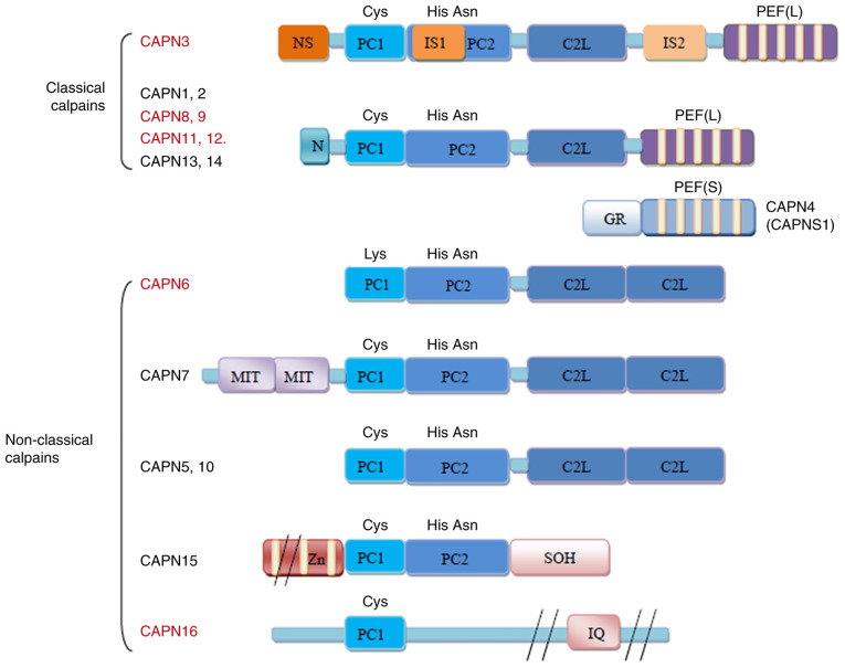
Figure 1.
Structure of the human calpains. The calpains presented in red are predominantly expressed in specific tissues or organs, while those in black are more widely expressed. The major difference in the structure of CAPN3 is that it contains three additional insertion sequences, namely NS at the N-terminus, IS1 of PC2 and IS2 between CBSW/C2L and PEF (L). Small subunits are not present in CAPN3. CAPN3, calpain 3; GR, Gly-rich domain; MIT, microtubule-interacting and transport motif; Zn, zinc-finger motif; SOH, small optic lobes product homology domain; IQ, calmodulin-interacting motif; NS, N-terminus; IS1, insertion sequence 1; PC1, protease core domain 1; PEF, penta-EF-hand (E, E-helix; F, F-helix); CBSW/C2L, calpain-type β-sandwich.

