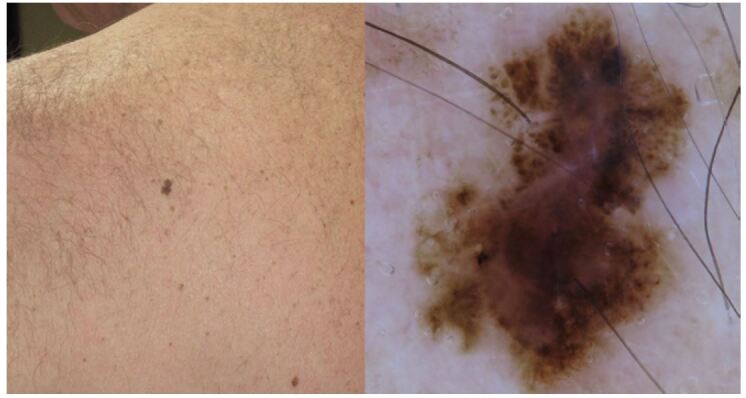
Figure 11.

Clinical and dermoscopic view of a small but largely atypical melanocytic lesion morphologically suggestive of melanoma. The patient’s wife reported the lesion as a long-standing stable macule. Again, the history was a confounding factor because the lesion was excised and diagnosed as an early invasive melanoma histopathologically.
