
Figure 9.
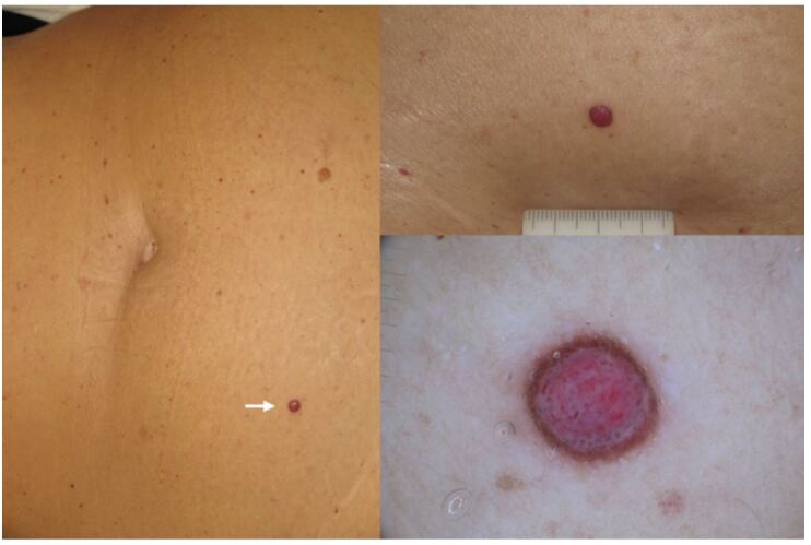
Clinical and dermoscopic view of a palpable lesion with symmetric distribution of colors and structures. Diagnosis of a benign lesion could not be made with confidence (several diagnostic options could be considered), therefore the lesion had to be excised with no monitoring. Subsequent histopathologic examination revealed a melanoma of 3.5 mm of thickness.