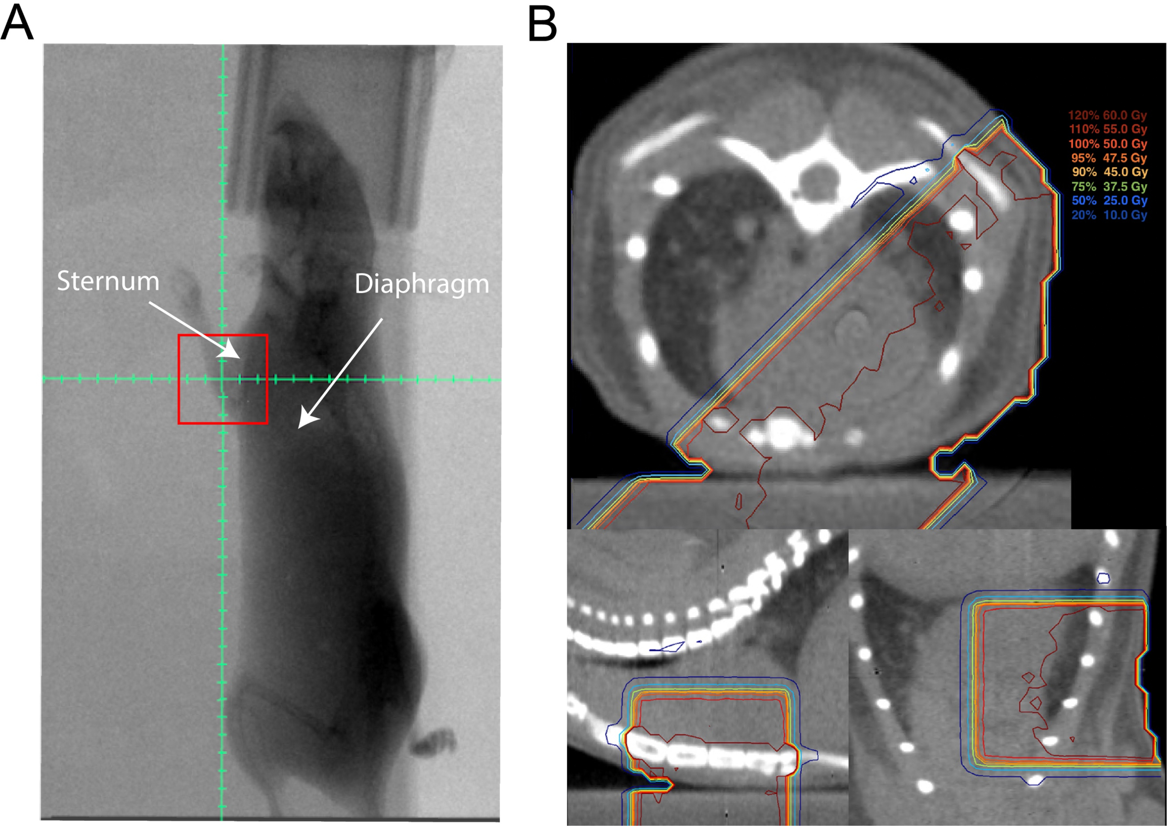
Figure 1.

Set-up and treatment plan for partial-heart radiation therapy. (A) Representative fluoroscopic image obtained at 315 degrees from right anterior oblique position with 10 × 10 mm treatment field (red square). Graticule with 2 mm tick marks shows isocenter, which was placed 2 mm anterior to sternum and 6–8 mm cranial to diaphragm. (B) Prescription isodose lines on a representative CT in the axial (top), sagittal (bottom left), and coronal (bottom right) planes showing prescription isodose lines covering part of the heart from opposed left posterior oblique and right anterior oblique treatment fields.
