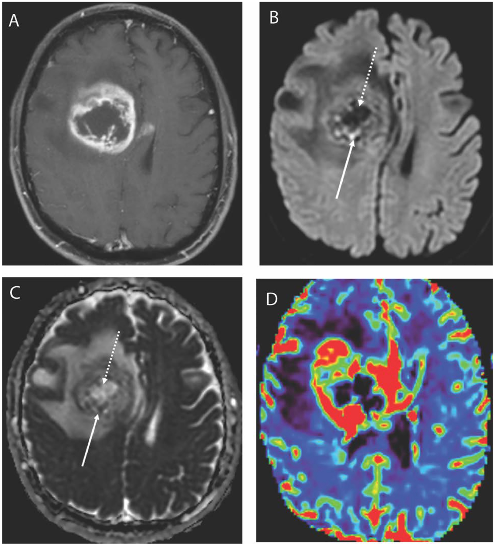
Figure 2.

Axial T1 weighted post contrast image (A) in a 67-year-old male with a glioblastoma demonstrates a large centrally necrotic mass in the right frontal lobe. DWI trace image (B) shows peripheral foci of restricted diffusion posteriorly suggestive of hypercellularity and the corresponding ADC map (C) confirms the presence of low ADC in these areas. Similarly, the central portion demonstrates facilitated diffusion with low signal on DWI trace image and elevated ADC.
