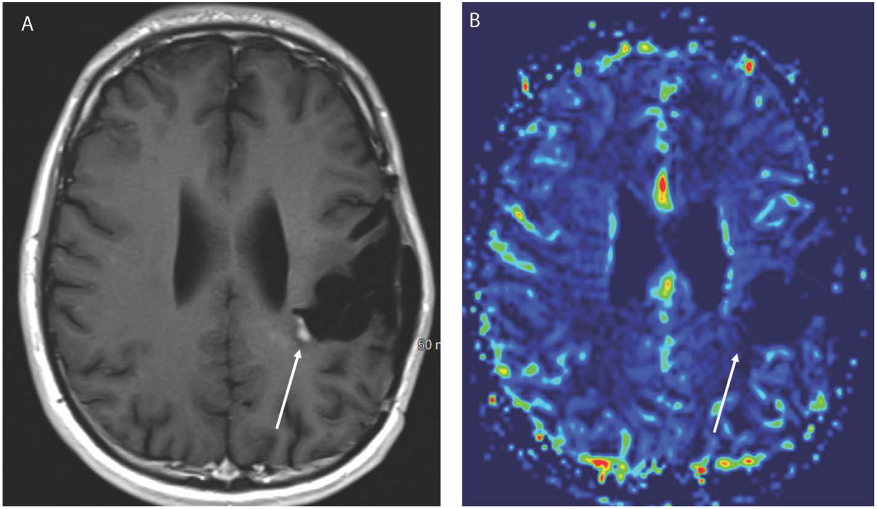
Figure 3.

Axial T1 weighted post contrast image (A) in a 27-year-old male with history of IDH mutated anaplastic astrocytoma after treatment with radiation and temozolomide 6 months earlier demonstrates a new focus of enhancement along the medial margin of the post-surgical cavity. The corresponding CBV map (B) from DSC perfusion, however, does not show elevated blood volume, and this was therefore considered to be pseudoprogression. Follow up imaging confirmed resolution of the enhancement.
