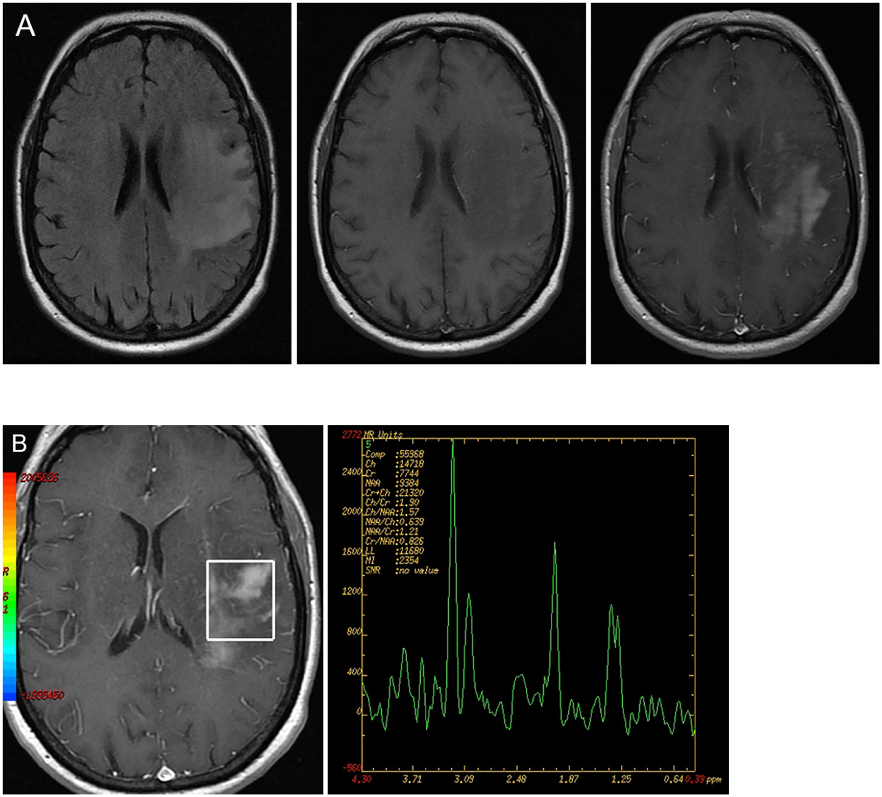
Figure 5A and 5B:

FLAIR hyperintense mass lesion showing predominantly T1 hypointense signal on the pre-contrast T1 weighted image. Heterogenous contrast enhancement present in the post contrast image. Single voxel MRS evaluation shows gross elevation of the choline peak and relatively diminished NAA peak (third from left) consistent with a neoplastic process, in this case of glioblastoma. (Image courtesy Prof. Mohannad Ibrahim, Michigan Medicine)
