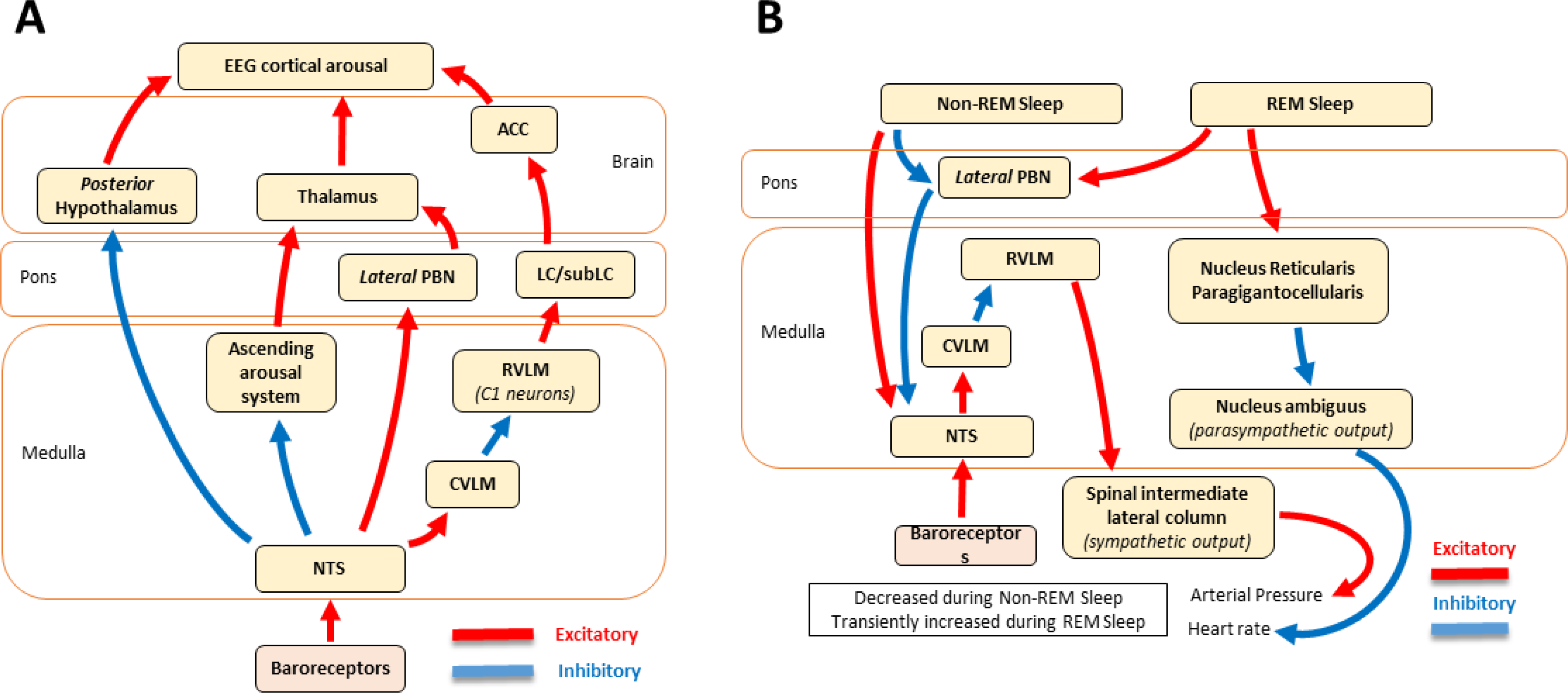
FIGURE 11.

The reciprocal influence between cardiovascular function and sleep. Arrows represent excitatory (red) and inhibitory (blue) functional influences that occur through monosynaptic or polysynaptic anatomical connections and relay nuclei (e.g., thalamus). Panel A: Oversimplified view of major neural structures involved in baroreceptor modulation of arousal. NTS projections to the ascending arousal system, posterior hypothalamus, and CVLM/RVLM reduce arousal and prompt sleep, whereas NTS projections to the PBN pathways promote arousal. Panel B: Sleep influences cardiovascular function. Non-REM sleep disinhibits the NTS, and as a result, decreases sympathetic output at the spinal lateral column, which enhances baroreflexes and reduces arterial pressure. In contrast, REM sleep reduces the parasympathetic output by inhibiting the nucleus ambiguus, which dampens baroreflexes and leads to a transient increase in AP and heart rate. ACC, anterior cingulate cortex; LC/subLC, locus coeruleus/locus subcoeruleus; Lateral PBN, lateral parabrachial nucleus; RVLM, rostral ventrolateral medulla; CVLM, caudal ventrolateral medulla; NTS, nucleus tractus solitarious. Lateral PBN and LC are part of the ascending reticular activating system.
