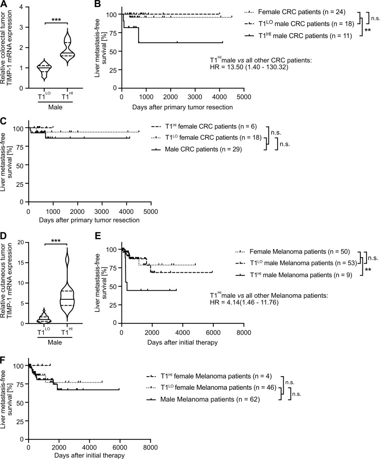
Figure 8.
Increased TIMP1 expression in tumors of male CRC or SKCM patients predicts early development of liver metastases. (A and D) TIMP1 mRNA expression in primary tumors of male CRC (A) or SKCM (D) patients separated by two-step cluster analysis into males with low (CRC: T1LO, n = 18; SKCM: T1LO, n = 53) or high (CRC: T1HI, n = 11; SKCM: T1HI, n = 9) TIMP1 expression. Student’s t test was employed for statistics. Medians (continuous lines) and interquartile ranges (dotted lines). (B and E) Probability of liver metastasis–free survival of male CRC (B) or SKCM (E) patients with high TIMP1 expression (CRC: T1HI, n = 11; SKCM: T1HI, n = 9) compared with all other patients (CRC: n = 42; SKCM: n = 103), respectively. The log-rank test was employed for statistics. HRs with 95% confidence intervals were determined by Cox regression analysis between T1HI males and all other patients. (C and F) Probability of liver metastasis–free survival of female CRC (C) or SKCM (F) patients with high TIMP1 expression (CRC: T1HI, n = 6; SKCM: T1HI, n = 4) compared with all other patients (CRC: n = 47; SKCM: n = 108). The log-rank test was employed for statistics. **, P ≤ 0.01; ***, P ≤ 0.001 (A–F).

