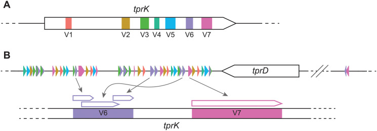
Fig 1. Anatomy of the tprK gene and mechanism of antigenic variation.
(A) tprK is composed of seven discrete variable regions (V1 –V7), color-coded here. (B) Flanking the tprD ORF (not to scale) are donor site segments used to generate unique V region sequences, again color-coded by their use in their respective variable regions. 51 of the known 53 donor sites are present in the 3’ flanking region. The segments used can be complete or partial, and vary in number for each V region sequence. They are combined through non-reciprocal segmented gene conversion to create antigenic variation.

