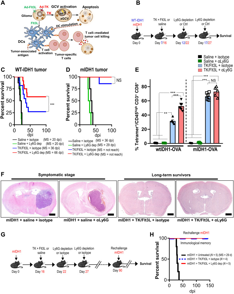
Fig. 1. mIDH1 tumor models have enhanced response to immune-stimulatory gene therapy irrespective of myeloid cell depletion.
(A) Schematic showing the mechanism by which TK/Flt3L gene therapy recruits and activates a tumor-specific T cell response. (B) Schematic illustrating the treatment strategy of the TK + Flt3L gene therapy in combination with Ly6G depletion in wtIDH1 or mIDH1 tumor–bearing mice. WT, wild type. (C and D) Kaplan-Meier survival curves of implanted mice-bearing wtIDH1 or mIDH1 tumors, treated with TK + Flt3L, Ly6G depletion, or combination therapy. dpi, days postimplantation; NS, not significant. (E) Tumor-specific CD8 T cell frequency within the TME of wtIDH1 or mIDH1 tumors treated with TK + Flt3L, Ly6G depletion, or combination therapy was analyzed by staining for SIINFEKL-Kb tetramers. (F) Hematoxylin and eosin staining of brain sections from mIDH1 tumors treated with saline, αLy6G, TK/Flt3L, and TK/Flt3L + αLy6G. (G) Schematic illustrating the treatment strategy of the TK + Flt3L gene therapy in combination with Ly6G depletion in mIDH1 tumor–bearing mice. Most mIDH1 tumor–bearing mice (90%) survived long term in response to TK/Flt3L gene therapy regardless of Ly6G depletion. The long-term survived mice were rechallenged at day 90 after implantation. (H) Kaplan-Meier survival plot for rechallenged long-term survivors from mIDH1 + TK/Flt3L + isotype (N = 4), mIDH1 + TK/Flt3L + Ly6G dep (N = 5), or control (mIDH1 + untreated) (N = 5). Data were analyzed using the log-rank (Mantel-Cox) test. Scale bars, 1 mm. **P < 0.01 and ***P < 0.005, One-way analysis of variance (ANOVA).

