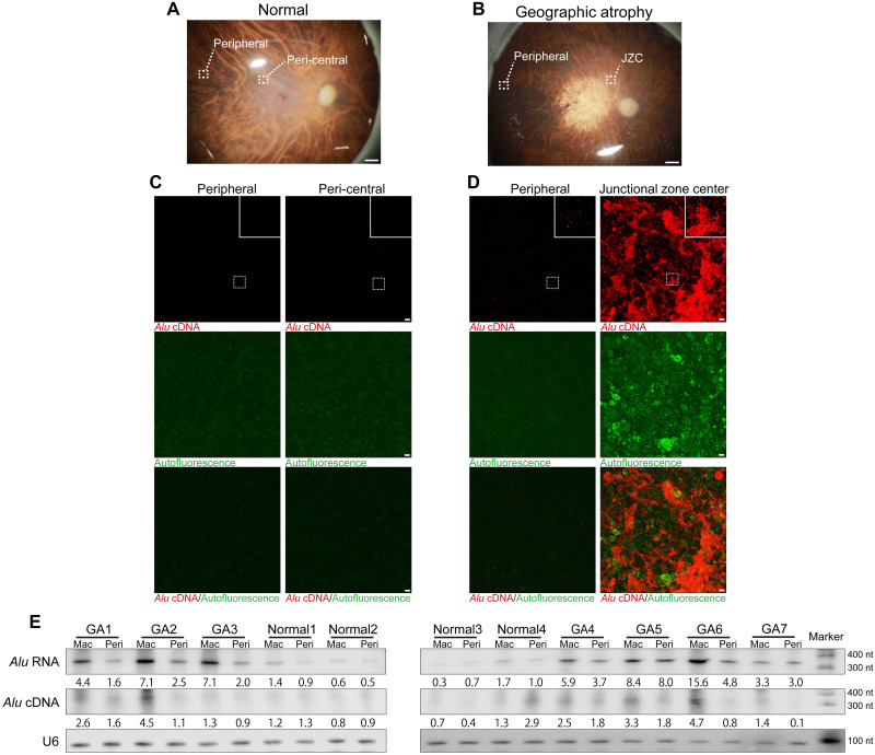
Fig. 1. Endogenous Alu cDNA in RPE of human geographic atrophy.
(A and B) Photographs of normal human donor eye retina illustrating peripheral and peri-central areas (A) and geographic atrophy (GA) retina illustrating peripheral and junctional zone center (JZC) areas (B). Scale bars, 1 mm. (C and D) In situ hybridization of RPE whole mounts with Alu cDNA–specific probes in peripheral and peri-central areas of normal eyes (C) and in peripheral and JZC areas of GA eyes (D). Insets show higher magnification. Red, Alu cDNA; green, autofluorescence. Scale bars, 10 μm. (E) Northern blotting of Alu RNA and equator blotting of Alu cDNA in macular (Mac) and peripheral (Peri) RPE of human GA (n = 7) and normal (n = 4) eyes. Densitometry of the bands corresponding to Alu RNA and Alu cDNA normalized to loading control (U6) with the mean densitometry ratio of macular RPE of normal eyes set to 1.0.

