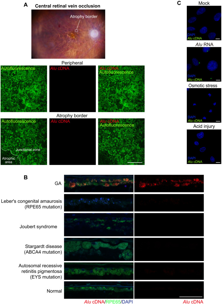
Fig. 2. Absence of endogenous Alu cDNA in human retinal diseases other than GA.
(A) Ex vivo fundus photograph of a human eye with RPE atrophy that developed subsequent to treatment of central retinal vein occlusion with anti-angiogenic drugs. In situ hybridization of RPE whole mounts showing no detectable endogenous Alu cDNA in peripheral RPE or at the border of the atrophic RPE. Scale bars, 200 μm. Red, Alu cDNA; green, autofluorescence of RPE cells. (B) Abundant endogenous Alu cDNA detected in the RPE of a human GA eye but not in the RPE of human eyes with Leber congenital amaurosis, Joubert syndrome, Stargardt macular dystrophy, or autosomal recessive retinitis pigmentosa. Red, Alu cDNA; green, RPE65; blue, 4′,6-diamidino-2-phenylindole (DAPI). Scale bars, 50 μm. (C) In situ hybridization of Alu cDNA formation in primary human RPE cells subjected to Alu RNA transfection, osmotic stress (distilled H2O), or acid injury (hydrochloric acid; HCl, pH 4.0 medium). Alu cDNA, green; DAPI, blue. Scale bars, 10 μm.

