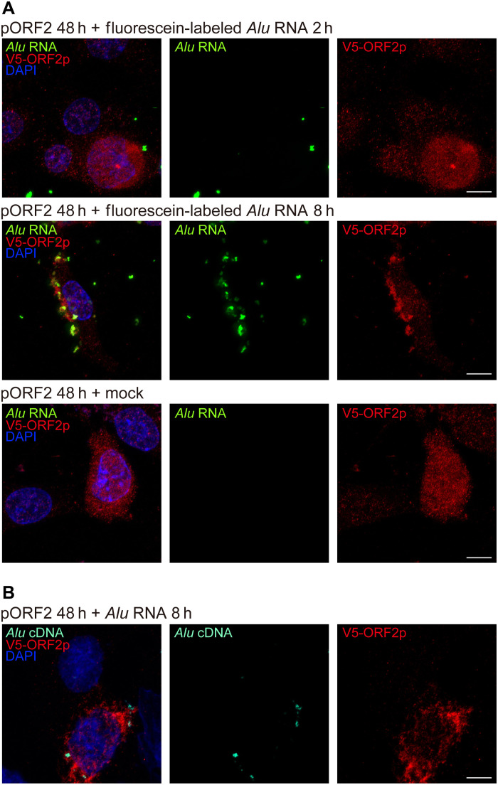
Fig. 5. Colocalization of L1 ORF2p, Alu RNA, and Alu cDNA.
(A) Fluorescence imaging of O. palustris RPE cells coexpressing ORF2p-V5 (detected by anti-V5 antibody, red) and fluorescein-labeled Alu RNA (transfected 48 hours after V5-ORF2 transfection, green). Images were acquired 2 and 8 hours after Alu RNA transfection. Note the diffuse localization of ORF2p-V5 and punctate foci of fluorescein-Alu RNA after 2 hours and cytoplasmic colocalization of multiple punctate foci 8 hours after Alu RNA transfection. ORF2p-V5 localization remained diffuse in the absence of transfected fluorescein-Alu RNA (mock, bottom). (B) In situ hybridization of O. palustris RPE cells shows coexpression of V5-ORF2 (red) and Alu cDNA (teal), detected by an Alu cDNA specific probe (48 hours after V5-ORF2 transfection and 8 hours after Alu RNA transfection). Note the cytoplasmic colocalization of ORF2p-V5 (red) and Alu cDNA at 8 hours after Alu RNA transfection. DAPI, blue. Scale bars, 10 μm.

