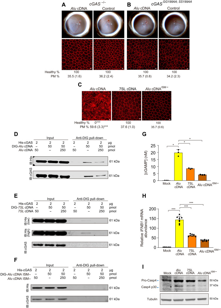
Fig. 7. Alu cDNA induces RPE degeneration via cGAS.
(A to C) Fundus photographs (top) and corresponding representative RPE sheet micrographs (bottom) of mice. Scale bars, 10 μm. Binary and morphometric quantification of RPE degeneration are shown. ***P < 0.001, Fisher’s exact test for binary; two-tailed t test for morphometry. PM, polymegethism [mean (SEM)]. RPE morphology after Alu cDNA or control administration in cGAS−/− mice (n = 6 to 8) (A) and catalytically null cGASGG198AA; SS199AA mice (n = 6) (B). (C) RPE morphology in WT mice after administration of Alu cDNA, 7SL cDNA, or a mutant form of Alu cDNA lacking a guanosine-rich ISM (Alu cDNAISM−). n = 6 to 8. (D to F) Immunoblotting of recombinant HIS-tagged cGAS using anti-HIS and anti-cGAS antibodies before (input) and after pull-down with anti–digoxigenin (DIG) antibody in the presence or absence of DIG-labeled or unlabeled competitor Alu cDNA (D), DIG-labeled or unlabeled competitor 7SL cDNA (E), or DIG-labeled or unlabeled competitor Alu cDNA lacking an ISM (Alu cDNAISM−) (F). Low- and high-exposure blots are presented in (E). (G) Enzyme-linked immunosorbent assay quantification of cGAMP production by recombinant cGAS in the presence of Alu cDNA, 7SL cDNA, or Alu cDNAISM−. (H) qRT-PCR quantification of IFNB1 mRNA in human RPE cells transfected with Alu cDNA, 7SL cDNA, or Alu cDNAISM−. *P < 0.05, ***P < 0.001, Mann-Whitney U test. The error bars in (G) and (H) represent the means ± SEM. (I) Immunoblots for pro-Casp4 and Casp4 p30 of human RPE cells transfected with Alu cDNA, 7SL cDNA, or Alu cDNAISM−.

