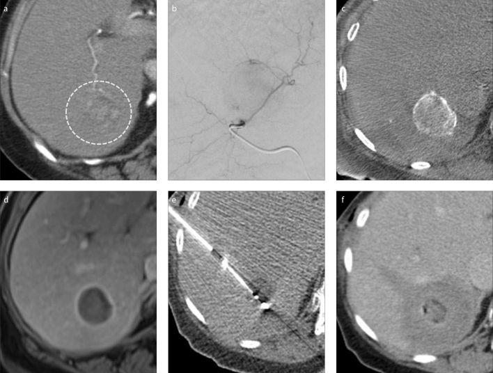
Figure. a–f.
Preprocedural axial CT (a) shows a 3.5 cm arterialized hepatocellular carcinoma (HCC) (circle). Digital subtraction angiogram (b) indicates the treatment position for transarterial chemoembolization (TACE). Unenhanced cone beam CT performed directly after TACE (c) shows contrast being entrapped between the beads in the target lesion. Follow-up MRI at 1 month (d) shows the lesion to be devascularized. In panel (e), the HCC had shrunk prior to treatment with microwave ablation and the hypoattenuating appearance post TACE helped allow visualization. Immediate post-procedural CT (f) shows an augmented ablation zone following TACE, with an appropriate margin. The patient remains disease-free at long-term follow-up.

