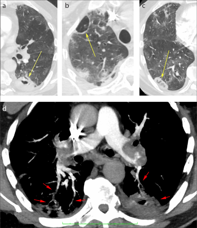
Figure. a–d.

Axial chest CT images showing two cavitary lesions, one with thick walls on the left lung (a, yellow arrow), and the other with thin walls on the right lung (b, yellow arrow). A reversed halo sign in the right lung is visible (c, yellow arrow). Pulmonary angio-CT image (d) demonstrating bilateral large emboli in the pulmonary arteries, irregular and tortuous small peripheral vessels (red arrows) related to the reversed halo sign in the right lung, and the cavitary lesion on the left lung.
