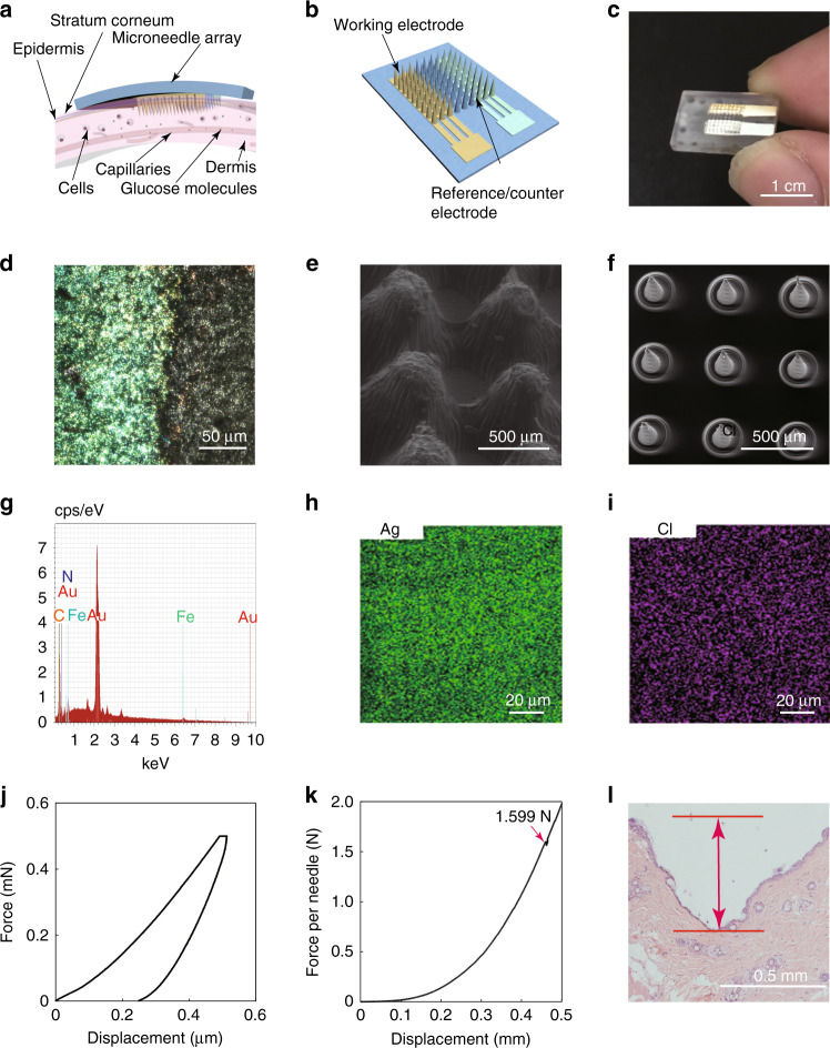
Fig. 1. Overall scheme and materials characterization of the microneedle biosensing device.
a A schematic illustration of the microneedle array inserted into the dermis of the skin and interstitial fluid. b A schematic illustration of the microneedle array. c A camera image of the microneedle-based electrochemical sensor. d An optical image of the Au electrode after the deposition of Prussian blue. e An SEM image of 3D printed cone-shaped microneedles with a base diameter of 400 µm and a height of 1.5 mm fabricated by using MoonRay (SprintRay Technology Ltd., China) and clear light-sensitive resin. f An SEM image of 3D printed cone-shaped microneedles with a base diameter of 200 µm and a height of 500 µm fabricated by using an S140 machine and biocompatible light-sensitive resin (BMF Precision Technology Ltd., China). g An EDS point analysis of the working electrode in the part of a microneedle. h, i EDS mapping of the Ag/AgCl electrode in part of a microneedle. j The load–displacement curve on a microneedle by an in situ nanomechanical test system. k Compression test on the microneedle array by a universal material testing machine. l An optical image of the pierced skin with staining after removing the microneedle array

