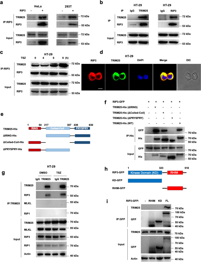
Fig. 1. RIP3 interacts with TRIM25.
a FLAG-tagged RIP3 was transfected into HeLa and HEK293T cells, followed by immunoprecipitation with the RIP3 antibody and immunoblotting analysis with TRIM25 and RIP3 antibodies, respectively. b Immunoprecipitation analysis with TRIM25 or RIP3 antibodies in HT-29 cells. c Co-immunoprecipitation analysis of RIP3 and TRIM25 by treatment with TNF/Z-VAD/Smac-mimetic (TSZ) stimuli for indicated hours in HT-29 cells. d RIP3 colocalized with TRIM25 in the cytoplasm of HT-29 cells. Endogenous RIP3 and TRIM25 were stained by anti-RIP3 (red) and anti-TRIM25 (green) antibodies, respectively. Scale bar, 10 µm. e Schematic of TRIM25 and different truncations. f His-tagged TRIM25 or its mutants and GFP-tagged RIP3 were co-transfected into HeLa cells. The cell lysates were immunoprecipitated with an anti-His antibody and then immunoblotted with the indicated antibodies. g HT-29 cells were primed with TSZ for 6 h, followed by immunoprecipitation with anti-TRIM25 antibody and probed with the indicated antibodies. h Schematic of RIP3 and different truncations. i GFP-tagged RIP3 or its mutants were transfected into HEK293T cells. The cell lysates were immunoprecipitated with an anti-GFP antibody and then immunoblotted with the indicated antibodies.

