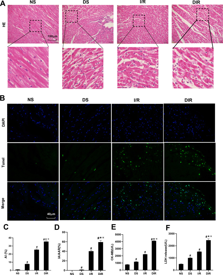
Fig. 1. DS, I/R, and DIR rat models construction and verification.
A Histopathological changes of myocardium were detected by HE staining. B, C The TUNEL staining for cell apoptosis rates were detected in different rat groups. D The myocardial infarction area was detected and expressed by AI/AAR (%). E CK-MB in serum was detected. F LDH in serum was detected. Results are presented by means ± SD. n = 8 per group. #P < 0.05 compared with the NS group. *P < 0.05 compared with the DS group. ※P < 0.05 compared with the I/R group.

