Figure 1.
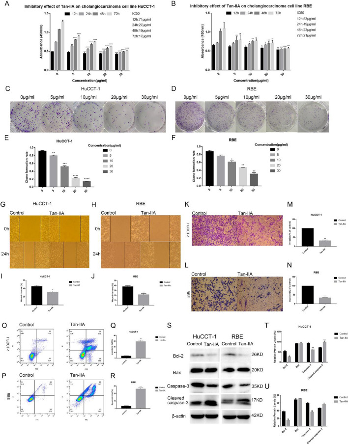
Cytotoxic effect of Tan-IIA on Cholangiocarcinoma cells. (A, B) Different concentrations (0, 5, 10, 20, and 30 µg/ml) of Tan-IIA were co-cultured with Cholangiocarcinoma cells for 12, 24, 48, and 72 h, and then the cytotoxic effect of Tan-IIA on Cholangiocarcinoma cells were detected by the CCK8 method. Cell viability histogram was made according to the formula (%) = [(experimental group OD value) − (blank group OD value)]/[(control group OD value) − (blank group OD value)] × 100%, and IC50 values were calculated for each time period. (C–F) Plate clone formation experiments were performed, Cholangiocarcinoma cells were co-cultured with Tan-IIA at concentrations of (0, 5, 10, 20, and 30 µg/mL) for two weeks and the number of clones was counted. Tan-IIA inhibits the migration and invasion of Cholangiocarcinoma cells. (G–J) Tan-IIA (24 h IC50 concentration) was co-cultured with Cholangiocarcinoma cells, then after scratching the cell layer for 24 h, cell migration by wound healing assay was determined. (K–N) The effect of Tan-IIA on the invasive ability of Cholangiocarcinoma cells was measured by the Transwell method. After crystalline violet staining, cell images were taken with an inverted microscope. Tan-IIA induced apoptosis of Cholangiocarcinoma cells. (O–R) The IC50 concentration for 24 h of Tan-IIA was selected and co-cultured with Cholangiocarcinoma cells for 24 h. Annexin V-FITC/PI double staining was used to detect apoptosis. (S–U) Western blotting to detect the effect of Tan-IIA on the expression of Bax, Bcl-2, as well as caspase-3 and cleaved caspase-3. Compared with control, *p < 0.05; **p < 0.01; ***p < 0.001; ****p < 0.0001.

