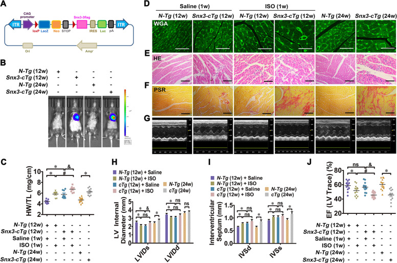
Fig. 2. Cardiac-specific Snx3 transgene led to myocardial injury in mice.
We investigated the changes in cardiac structure and function in Snx3-cTg mice at 12 and 24 weeks. Besides, Snx3-cTg mice (at 11 weeks) were injected with 3 mg/kg/d ISO (s.c.) or an equal volume of sterile normal saline for one week. A The scheme of Snx3-cTg mice construction strategy was shown. B The protein level of SNX3 was detected by live imaging. C The HW/TL ratio was calculated. D–F WGA staining (Scale bar: 50 μm), HE staining (Scale bar: 100 μm) and PSR staining (Scale bar: 100 μm)-stained cross-sections of heart tissues were shown. G The representative echocardiographic graphs were shown. H–J The echocardiographic parameters (LVID, IVS, and EF) were measured. Representative images of five independent experiments were presented. The data were shown as the means ± SEM. *P < 0.05 vs. N-Tg (12w) + Saline or N-Tg (24w) group, #P < 0.05 vs.. N-Tg (12w) + ISO group, &P < 0.05 vs. Snx3-cTg (12w) + Saline group. n = 12 per N-Tg (12w) + Saline group, n = 11 per N-Tg (12w) + ISO group, n = 12 per Snx3-cTg (12w) + Saline group, n = 12 per Snx3-cTg (12w) + ISO group, n = 10 per N-Tg (24w) group, n = 9 per Snx3-cTg (24w) group. EF ejection fraction, HE hematoxylin–eosin, HW/TL the heart weight to the tibia length ratio, ISO isoproterenol, IVS interventricular septum, LVID left ventricular diameter, ns no statistical difference, N-Tg non-transgenic, PSR picric sirius red, s.c. subcutaneously, Snx3-cTg cardiac-specific Snx3 transgenic mice, WGA wheat germ agglutinin, 1w 1 week, 12w 12 weeks, 24w 24 weeks. See also Supplementary Figs. S5–S7.

