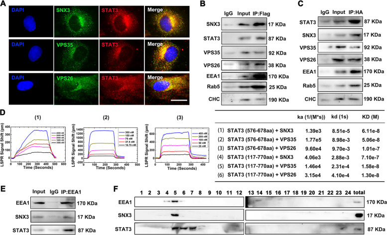
Fig. 3. SNX3–retromer directly interacted with STAT3 at early endosomes in vivo and in vitro.
A NRCMs were infected with Ad-STAT3 (HA-tagged) and were measured by IF staining using confocal microscopy (Scale bar: 25 μm). Representative images of five independent experiments were presented. NRCMs were infected with Ad-SNX3 (Flag-tagged) or Ad-STAT3 (HA-tagged) and were precipitated by anti-Flag (B) or anti-HA (C). D The binding curves of SNX3-retromer and truncated STAT3 protein (576-678aa) were measured by LSPR analysis, and the ka, kd, and KD values for STAT3 (576–678aa, 117–720aa), SNX3, VPS35, and VPS26 were calculated by TraceDrawer™. E NRCMs were precipitated by anti-EEA1 (a marker for early endosome) for SNX3 and STAT3 detection in co-IP assays. F The early endosome fraction was purified from NRCMs using density gradient centrifugation, and detected by western blot. n = 5. CHC clathrin heavy chain; co-IP co-immunoprecipitation, IP–MS immunoprecipitation-based mass spectrometry, LSPR localized surface plasmon resonance, NRCMs neonatal rat cardiomyocytes. See also Supplementary Figs. S8 and S9.

