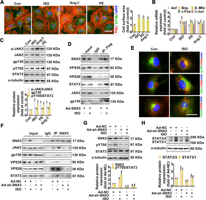
Fig. 4. SNX3–retromer acted as a platform for STAT3 activation in NRCMs.
NRCMs were treated with three hypertrophic stimulants, including ISO (10 μmol/L), Ang II (1 μmol/L), and PE (100 μmol/L), respectively, for the times indicated. A The cell surface area was measured by staining with anti-MLC2 antibody (green) and rhodamine-phalloidin (red) (Scale bar: 25 μm). B The mRNA levels of Anf, Bnp, β-Mhc, c-Fos, and c-Jun were determined by qPCR. C Western blot analysis was performed to detect the phosphorylated JAK2 (at tyrosine 1007 and 1008, p-JAK2) and STAT3 (at tyrosine 705, pY705), as well as the protein expression of JAK2, gp130 and STAT3. D NRCMs infected with Ad-SNX3 (Flag-tagged) were treated with ISO for 1 h and were precipitated by anti-Flag antibody, followed by co-IP assays. E The intracellular co-localization of SNX3 and gp130, JAK2, STAT3 in ISO-treated NRCMs was determined using confocal microscopy (Scale bar: 25 μm). NRCMs were infected with Ad-sh-SNX3 followed by incubation with ISO (10 μmol/L for 1 h), were precipitated by anti-SNX3 in co-IP assays (F), were detected the phosphorylated STAT3 (pY705), the protein expression of SNX3 and STAT3 (G), and were examined the protein expression of STAT3/3 homodimer or STAT3/1 heterodimer (H). Representative images of five independent experiments were presented. The data were shown as the means ± SEM. *P < 0.05 vs. control or Ad-NC group, #P < 0.05 vs.. Ad-NC + ISO group, n = 5. Ang II, angiotensin II; co-IP, co-immunoprecipitation, IF immunofluorescence, ISO isoproterenol, MLC2 myosin light chain 2, NC negative control, NRCMs neonatal rat cardiomyocytes, PE phenylephrine, qPCR quantitative polymerase chain reaction, 1h, 1 h. See also Supplementary Figs. S10 and S11A.

