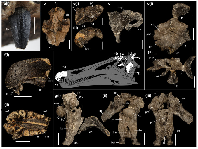
Figure 4.
Cranial material of Riparovenator milnerae. (a) Close up of holotype in situ RpmVII (IWCMS 2014.95.6), in labial view; (b) referred posterior nasal fragment (IWCMS 2014.95.7) in dorsal view; (c) holotype left preorbital fragment (IWCMS 2014.96.3) in (i) lateral and (ii) anterodorsal view; (d) holotype right laterosphenoid (IWCMS 2014.96.2) in lateral view; (e) holotype skull roof and associated left laterosphenoid (IWCMS 2014.96.1) in (i) dorsal and (ii) left lateral views; (f) holotype premaxillary bodies (IWCMS 2014.95.6) in (i) left lateral and (ii) ventral views; (g) holotype basicranium (IWCMS 2020.448.1) in (i) right lateral (with fractured cultriform process IWCMS 2020.448.2), (ii) posterior and (ii) anterior views. bo basioccipital, bs basisphenoid, bpt basipterygoid process, bsr basisphenoid recess, cap capitate process of the laterosphenoid, cp cultriform process, exo exoccipital, f frontal, fl fluting, fm foramen magnum, fp frontal process, lac lacrimal, ls laterosphenoid, mn maxillary notch, mr median ridge, oc occipital condyle, os orbitosphenoid, p parietal, plp posterolateral process, pop postorbital process, pm(n) premaxillary tooth/alveolus (tooth position), prf prefrontal, pro prootic, sc sagittal crest, scr subcondylar recess, so supraoccipital, ssr subsellar recess, stf supratemporal fossa, vp ventral process of the prefrontal. Skull reconstruction credit: Dan Folkes (CC-BY 4.0). Scale bars (a): 5 mm; (b–d): 20 mm; (e–g): 50 mm.

