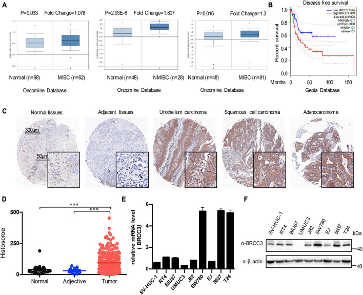
FIGURE 1.
Expression of BRCC3 in bladder cancer. (A) Data form Oncomine database. Three microarray datasets exhibited obvious upregulation of BRCC3 expression in muscle-invading bladder cancer tissue compared to normal. (B) Disease-free survival time analysis of data from the GEPIA database. The p-value is indicated. (C) Immunohistochemical staining for BRCC3 in tissue microarrays containing cancer tissue samples, adjacent tissue samples and normal bladder tissue samples from patients with chronic cystitis or a healthy urinary tract. Representative staining images for different pathological types of bladder cancer including uro-endothelial carcinoma, squamous cell carcinomas and adenocarcinoma are shown. (D) Tissue microarray data analysis of BRCC3 expression in 188 bladder cancer tissue samples, 12 corresponding adjacent tissue samples and 16 normal bladder tissue samples (including 8 chronic cystitis tissue samples and 8 healthy bladder tissue samples collected at autopsy). (E) The mRNA expression levels of BRCC3 in SV-HUC-1 and 8 bladder cancer cell lines. (F) The protein expression levels of BRCC3 in SV-HUC-1 and 8 bladder cancer cell lines. *P < 0.05, **P < 0.001, and ***P < 0.001 compared with controls.

