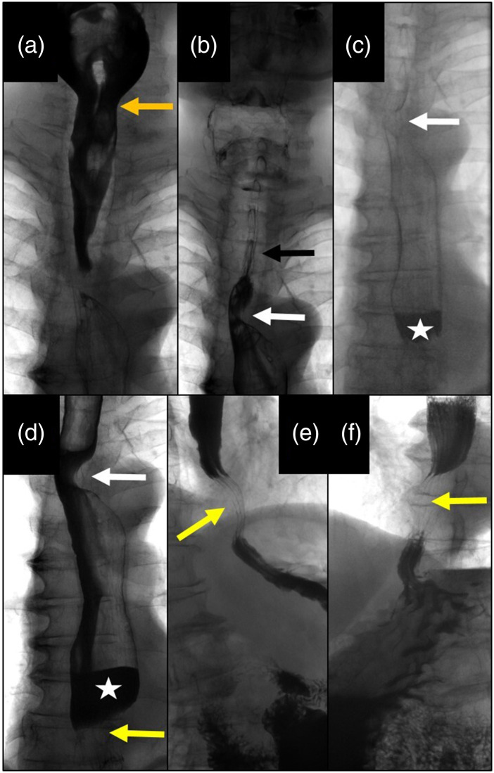
Fig. 1.
Barium swallow images: (a) Normal pharyngeal phase with regular opening of the upper sphincter. (b) Normal barium passage through the upper esophagus with a regular mucosal surface (black arrow): lateral impression due to an accessory arteria lusoria (white arrow). (c) Dilation of the distal esophagus with slow passage of barium and formation of an air-barium level (white asterisk). (d) Distal esophagus dilated with air-barium level (asterisk) due to aortic compression (lower arrow); note the impression of the arteria lusoria (upper arrow). (e) Right lateral view of the aortic impression of the distal esophagus (arrow). (f) Left lateral view of the esophageal compression 3 cm above the diaphragm due to the crossing aorta (arrow) with upstream barium congestion.

