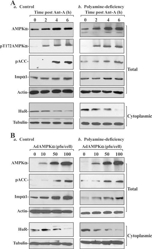
Figure 3. Effect of stimulation of AMPKα by treatment with its chemical activator antimycin A (Ant-A) and ectopic expression of WT AMPKα gene on cytoplasmic levels of HuR in control and polyamine-deficient cells.

(A) Changes in levels of total and pAMPKα, pACC, Impα1 and HuR after treatment with Ant-A. (a) Control; (b) polyamine-deficient cells. IEC-6 cells were cultured in the control medium and a medium containing DFMO for 6 days and then exposed to Ant-A (5 μM). Whole-cell lysates were harvested at different times after administration of Ant-A, and total and cytoplasmic fractions were prepared for Western-blot analysis. Levels of AMPKα, pAMPKα, pACC, Impα1 and HuR were identified by using the specific antibodies, and equal loading was monitored by immunoblotting of β-actin in total proteins and β-tubulin in cytoplasmic proteins. Three experiments were performed and they showed similar results. (B) Changes in levels of AMPKα, pACC, Impα1 and HuR after overexpression of the AMPKα gene. (a) Control; (b) polyamine-deficient cells. After cells were grown in control cultures and cultures containing 5 mM DFMO for 4 days, they were infected with the adenoviral expression vector encoding the complete open reading frame of the human AMPKα cDNA (AdAMPKα) or adenoviral vector lacking AMPKα cDNA (Adnull) at an MOI (multiplicity of infection) of 10–100 pfu per cell. Whole-cell lysates were harvested 48 h after the infection in the presence or absence of DFMO, and levels of AMPKα, pACC, Impα1 and HuR were measured by Western-blot analysis. Three experiments were performed and they showed similar results.
