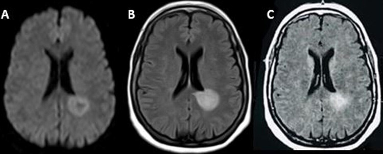
Figure 1. Brain magnetic resonance imaging (1.5 Tesla) showed two pseudo-nodular lesions of the peri-ventricular white matter, hyperintense in T2 and T2 flair, isointense in T1, with diffusion restriction, associated with other small lesions of the white matter on the left frontal level, right insular and bilateral occipital, enhanced after injection of gadolinium.
(A) Diffusion axial view; (B) T2 fluid-attenuated inversion recovery (FLAIR) axial view; (C) T1 axial view with gadolinium.

