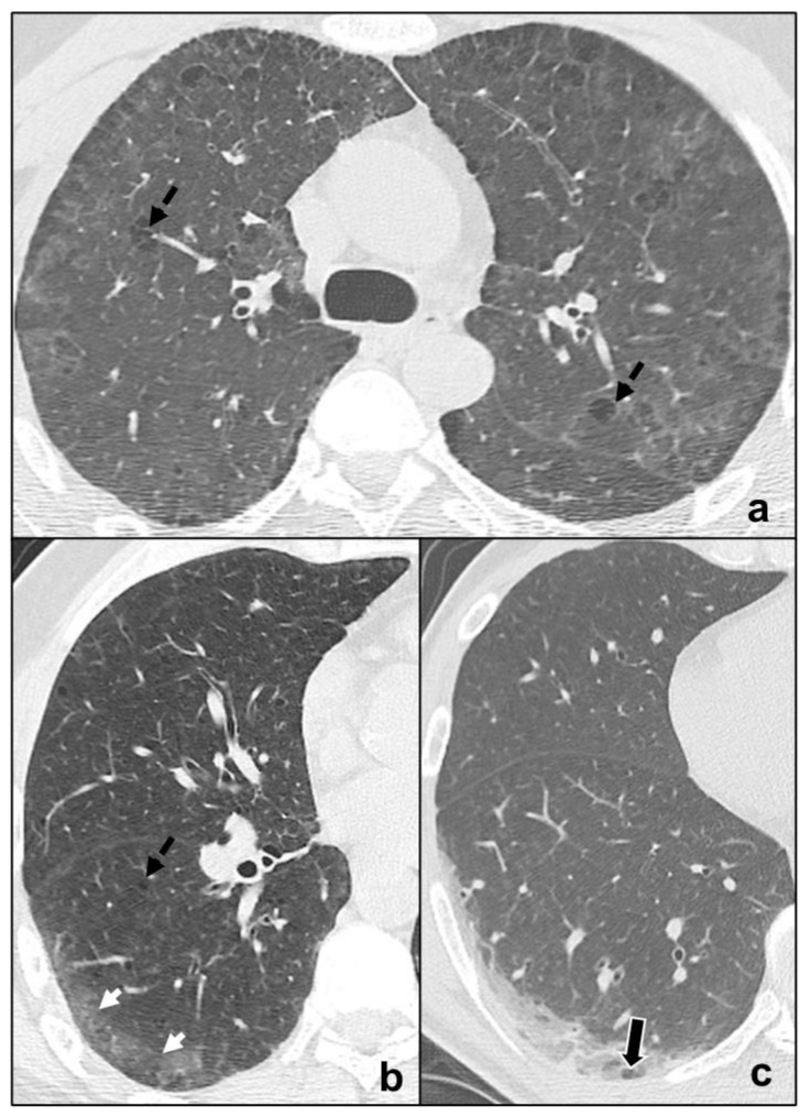
Figure 4.
Major IIPs: Smoking-Related IIPs. In (a), RB-ILD is characterized by bilateral widespread ground glass opacities, centrilobular nodules, and centrilobular (black dotted arrows), and subpleural emphysema; in (b,c), DIP shows subpleural ground-glass areas (white arrow in (b)) associated with reticular, thin opacities and centrilobular (black dotted arrows) emphysema, while in (c), perivascular cysts are evident (white-bordered black arrow in (c)).

