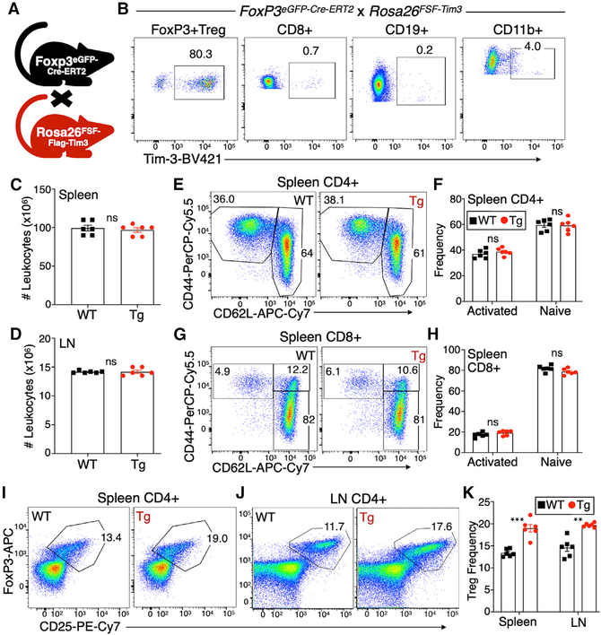
Figure 2. Increased Tim-3+ Treg cell frequency in peripheral lymphoid organs of FoxP3EGFP-Cre-ERT2 × FSF-Tim-3 mice.
(A) Depiction of the cross used to generate FoxP3EGFP-Cre-ERT2 × FSF-Tim-3 mice.
(B) Tim-3 transgene expression is restricted to CD4+ Treg cells when driven by the FoxP3-specific, tamoxifen-inducible Cre. Mice (both WT and Tg) were dosed on 5 consecutive days with tamoxifen, followed by 1 day of rest, before analysis. Shown is the analysis of splenic lymphocytes from representative animals.
(C and D) No change in the total number of splenocytes or lymph node cells isolated from WT versus Tim-3 Tg animals after tamoxifen treatment.
(E–H) Antigen-experienced and naive conventional CD4+ (E and F) and CD8+ (G and H) T cells from spleens of WT and Tim-3 Tg animals, based on CD44 and CD62L expression.
(I–K) Proportions of FoxP3+CD25+ Treg cells among CD4+ T cells in the spleen (I) and lymph nodes (J) of WT and Tim-3 Tg animals, quantified in (K).
Graphs show individual animals and mean ± SEM; two-tailed t test.

