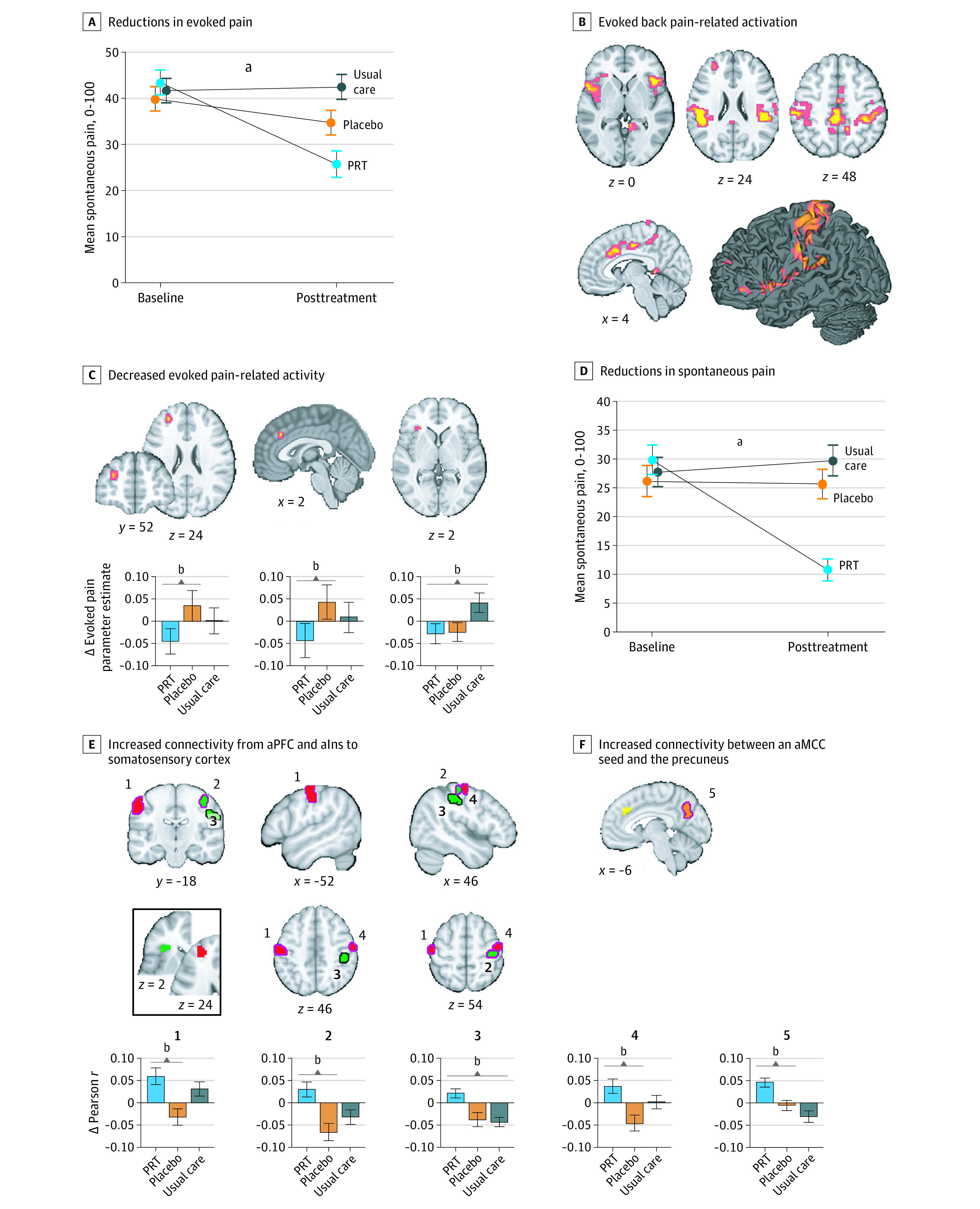
Figure 3. Effects of Treatment on Evoked and Spontaneous Back Pain and Related Brain Function.

A, Error bars show standard error. B, Coordinates and statistics for activations provided in eTable 7 in Supplement 2; analyses conducted within a mask of regions of interest; eFigure 1 in Supplement 2. C, Decreased evoked pain-related activity was observed in anterior midcingulate (aMCC) and anterior prefrontal regions for PRT vs placebo and left anterior insula for PRT vs usual care. D, Error bars show standard error. E, PRT vs control conditions increased aPFC-seeded (red clusters) and aIns-seeded (green clusters) connectivity with primary somatosensory cortex (permutation test, P < .05). Inset shows seed regions, derived from evoked pain analyses; magenta outlines, PRT vs placebo; black outlines, PRT vs usual care. F, PRT vs usual care increased connectivity between an aMCC seed (yellow; derived from evoked back pain analyses) and the precuneus (orange). Connectivity analyses were conducted within primary somatosensory cortex and medial default mode network masks.
aP < .001.
bP < .05.
