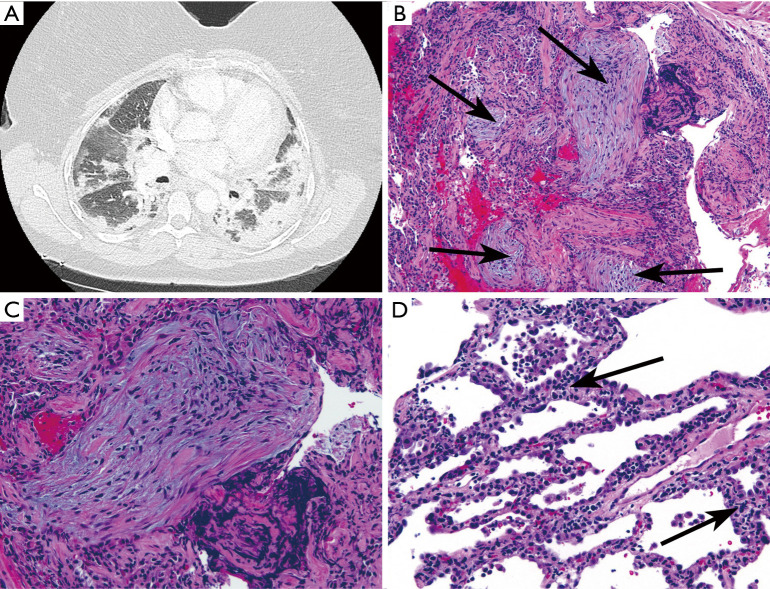
Figure 3.
Transbronchial biopsy from a woman in 7th decade of life with AS, positive for anti-Jo-1. (A) Chest CT showed bilateral peripheral and lower lobe predominant consolidative and ground-glass opacities interpreted as mixed OP/NSIP. (B) Transbronchial biopsy showing organizing pneumonia (arrows indicate fibroblast plugs; HE, ×100). (C) Fibroblast plug (Masson body) at higher magnification. (D) Diffuse interstitial chronic inflammation (arrows; HE, ×200). These findings could be interpreted as a combination of organizing pneumonia and NSIP (HE, ×200).

