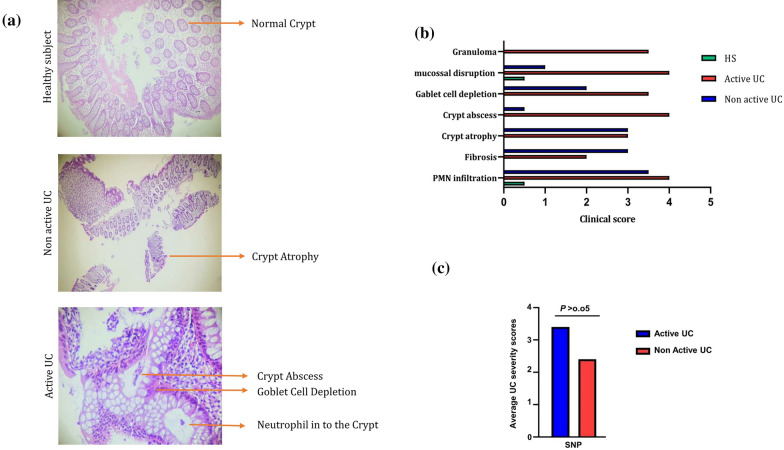
Fig. 1.
a H&E stained colon sections of HS, non-active UC, and active UC samples. The picture was taken using an Olympus D330 digital camera (Olympus, Tokyo, Japan) (×40 magnifications). b Damage scores ranged from 0 to 4, as described in the text. Scales were judged based on the number and extent of PMN infiltration, mucosal disruption, crypt abscess, crypt atrophy, goblet cell depletion, fibrosis, and granuloma. c There was no significant difference between the average UC severity scores of the SNP-positive and SNP-negative patients in the UC group. Data are presented as mean ± SD

