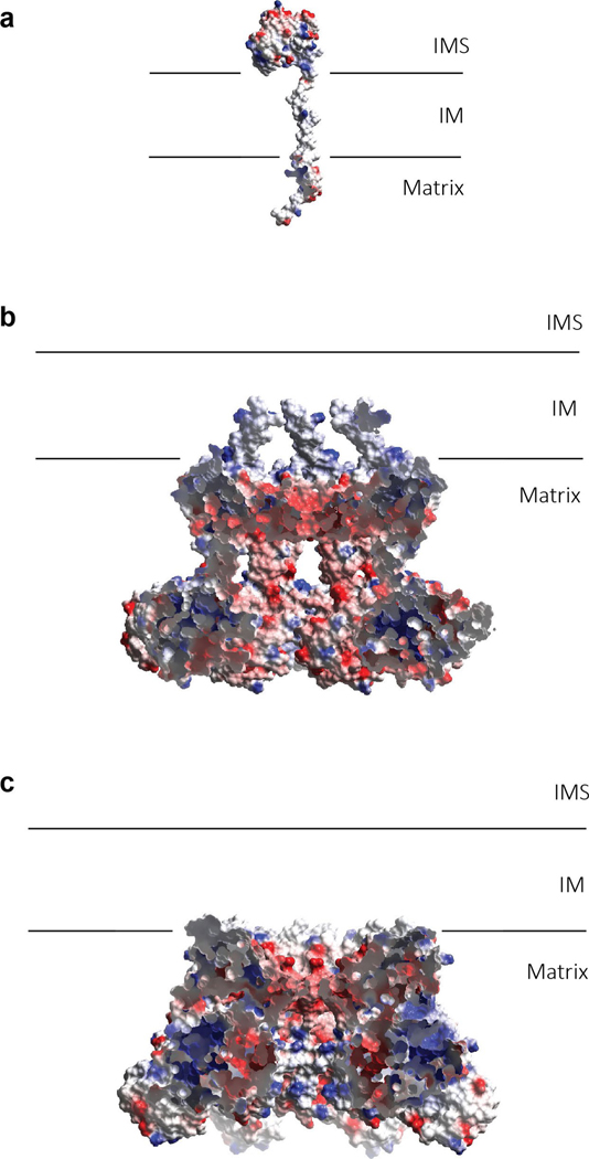
Extended Data Fig. 9 |. Electrostatic surface potential of substrate and the putative substrate-binding cavity of mBcs1 in different states.
a, Electrostatic potential surface for the ISP subunit. b, Electrostatic potential surface of the apo mBcs1. The front portion of the surface was cut away to reveal the interior surface potential of the putative substrate-binding cavity. c, Electrostatic potential surface of the mBcs1 bound with ATPγS. The front portion of the surface was cut away to reveal the interior surface potential of the putative substrate-binding cavity.

