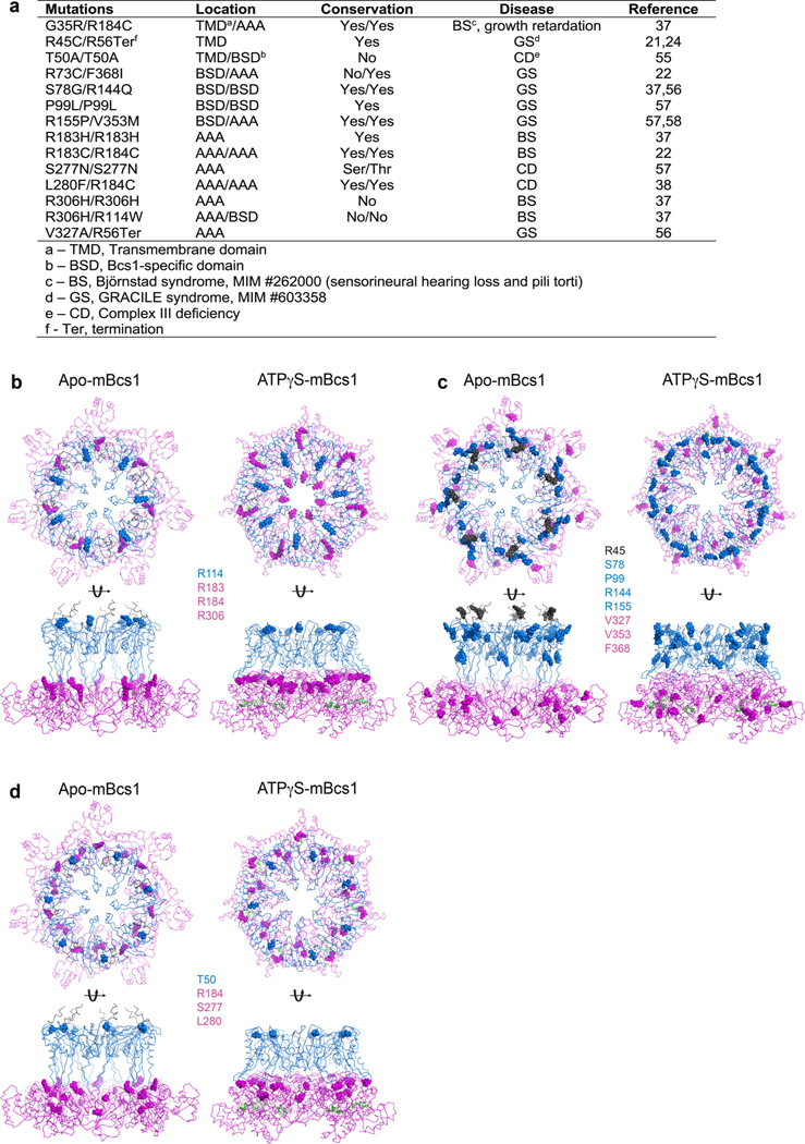
Extended Data Fig. 10 |. Pathogenic mutations of BCS1L.
a, Pathogenic mutations of BCS1L and their locations in the Bcs1 structure. b–d, Mapping of the mutations on ATPγS-bound and apo mBcs1 in two orthogonal orientations: top view (top) and side view (bottom). TM, Bcs1-specific and AAA regions are in black, blue and magenta ribbons, respectively. Mutations are showed in spheres. Mutations found in (b), Björnstad syndrome; (c) GRACILE syndrome and (d) Complex III deficiency.

