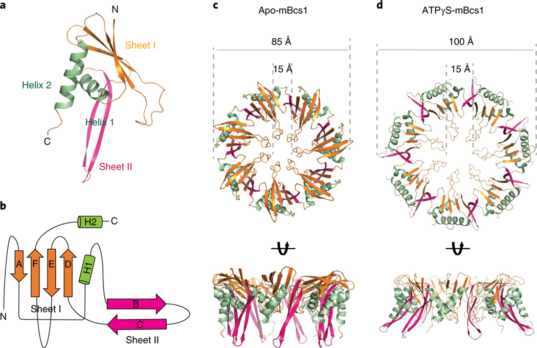
Fig. 2 |. Bcs1-specific domain of mBcs1.
a, Cartoon representation of the Bcs1-specific domain. The β-Sheet I and Sheet II are colored coral and magenta, respectively. The α-helices H1 and H2 are shown in green. b, Topological drawing showing the connectivity of the secondary structures of the Bcs1-specific domain. c, Heptameric association of the Bcs1-specific domains in the apo structure in two orthogonal views. The color code is the same as in a. d, Heptameric association of the Bcs1-specific domains in the ATPγS-bound structure.

