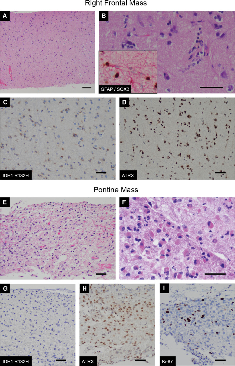
Fig. 2.
Genetically divergent multifocal glioma involving cerebrum and brainstem. A–D Biopsies of the cortical mass revealed a diffusely infiltrating glial neoplasm with perineuronal satelitosis and perinuclear clearing (A, 4×; B, 400×). The neoplastic cells expressed SOX2 (brown chromogen) and GFAP (red chromogen) (B inset, 200×), and harbored the IDH1 R132H onco-protein (C, 200×) with retained nuclear expression of ATRX (D, 200×). E–I Biopsies of the pontine mass revealed a diffusely infiltrating glial neoplasm with increased cellularity and prominent mini-gemistocytic cytomorphology (E, 200×; F, 400×). As opposed to the cortical mass, the pontine mass lacked the IDH1 R132H onco-protein (G, 200×) however nuclear expression of ATRX was similarly retained (H, 200×). Ki-67 index was increased in the pontine biopsy – a representative image is provided (I, 200×). (Scale bar = 50 um.)

