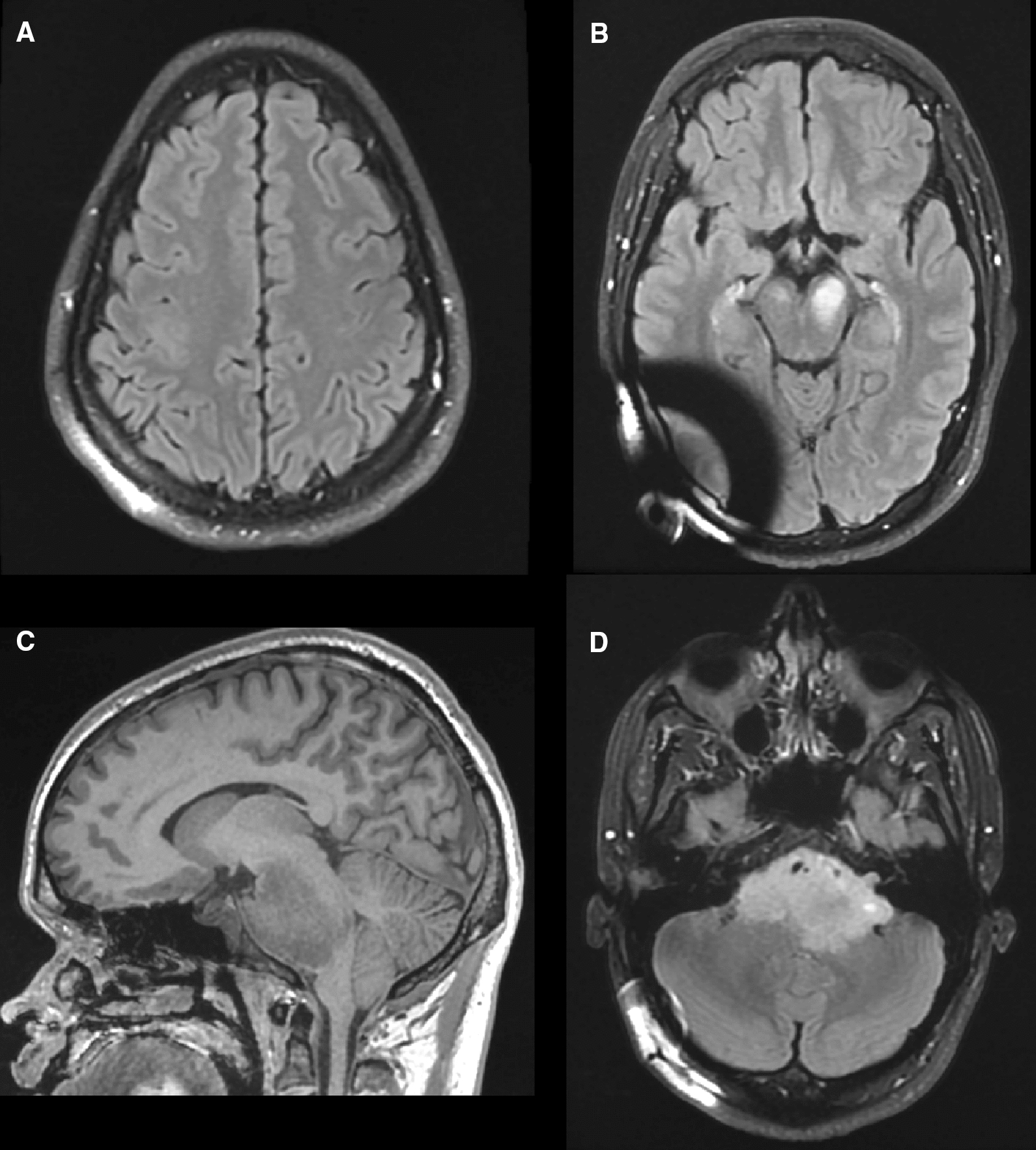
Fig. 3.

Follow up MRI following four cycles of PCV. A Hyperintense lesion of similar size and appearance in the precentral gyrus on axial T2-FLAIR. B Homogenous hyperintense lesions in the left and right cerebral peduncles on axial T2-FLAIR. C Large, primarily hypointense pontine lesion on sagittal T1 measuring 6.1 × 4.8 × 3.9 cm. Note herniation of the cerebellar tonsil. D Axial T2-FLAIR demonstrating new hyperintense foci along the left anterior base of the lesion that extends into the cerebellopontine cistern and internal auditory canal. Again, encasement of the basilar artery is noted but flow is patent
