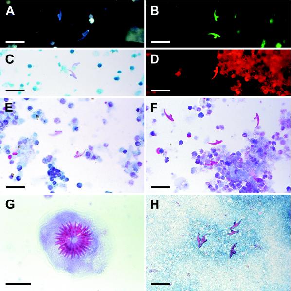
FIG. 1.
(A) Hydatid hooklets, methanol fixed, revealed by epifluorescence microscopy (excitation filter wavelength, 365 nm; long-pass filter wavelength, 420 nm); (B) hydatid hooklets on polycarbonate filter, methanol fixed, examined by epifluorescence microscopy (excitation, 405 nm; long pass, 495 nm); (C) hydatid hooklets revealed by Ziehl-Neelsen stain; (D) hydatid hooklet on polycarbonate filter revealed by Ziehl-Neelsen stain and epifluorescence microscopy (excitation, 546 nm; long pass, 590 nm); (E) hydatid hooklet revealed by trichrome stain; (F) hydatid hooklets revealed by Ryan stain; (G) hydatid protoscolex revealed by Ryan stain; (H) hydatid hooklets revealed by modified Baxby stain. Photomicrographs were shot on negative film, scanned into Kodak Photo CD disks, and processed with Adobe Photoshop version 4.0. Scale bars, 100 μm (G) and 30 μm (all other panels).

