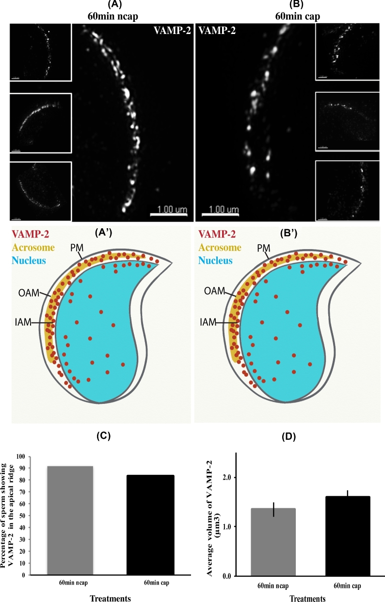
Figure 8.
VAMP2 detected by SR-SIM does not redistribute in sperm during capacitation. (A) Images of sperm incubated in noncapacitating medium (ncap) for 60 min. A punctate pattern of VAMP2 can be observed at the apical ridge of the sperm in the large image as well as three insets. (A΄) Schematic representation of (A). (B) Images of sperm incubated in capacitating medium (cap) for 60 min. A similar pattern of VAMP2 can be observed at the apical ridge of the sperm head when compared with (A). (B΄) Schematic representation of (B). The larger image and three insets show a similar result. (C) Images sperm at 60 in of incubation in capacitating medium and noncapacitating conditions collected using SR-SIM microscopy were quantified. The percentage of sperm with VAMP2 at the apical ridge was plotted. (D) The volume of VAMP2-positive fluorescence on sperm head was calculated. Images were collected at 60 min from sperm incubated in capacitating and noncapacitating medium. A total of 16 sperm were analyzed for each treatment and time point. No statistical difference was found.

