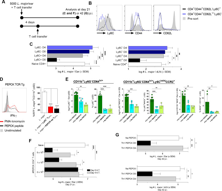
Fig 5. Circulating TEFF availability prior to niche establishment is required for L. major control.
(A-G) Naïve C57Bl/6 mice were infected with L. major and adoptively transferred immediately (C, E-G) or 4 days p.i. (C, F, and G)) with (C) 2.5–3x10⁶ Ly6C+CD44+CD62L-CD4+ or 5.5–6x10⁶ Ly6C-CD44+CD4+ FACS sorted polyclonal T cells derived from the blood, spleen, and dLNs of chronically infected C57Bl/6 mice or (E-G) in-vitro-generated Th1 PEPCK-specific GFP+ TcR Tg T cells. (B) Analysis of Ly6C+, CD44+, and CD62L- expression on polyclonal CD4+ T cells derived from chronically infected mice pre- and post-cell sorting. (C) Parasite loads in the ear dermis and ear dLNs following polyclonal CD4+ T cell transfer as determined by LDA at day 21 p.i.. (D) Representative histogram of IFN-γ+ Th1 PEPCK-specific in-vitro-generated T cells after PMA-Ionomycin, PEPCK peptide, or no stimulation (left panel) and comparison of %L. major-RFP in-vitro in bone marrow-derived monocytes by chronic mouse-derived Ly6C+CD44+CD62L-CD4+ TEFFs or in-vitro-generated Th1 PEPCK-specific GFP+ TcR Tg T cells (right panel).. (E) Analysis of the frequency and absolute number of the indicated popuations on day 4 p.i. and adoptive transfer of 4-5x10⁶ Na or Th1 primed PEPCK TcR Tg or Th1 primed SMARTA TcR Tg CD4+ T cells. (F) Parasite loads in the ear dermis following Th1 PEPCK-specific transfer of the indicated number of cells as determined by LDA at day 21 p.i.. (G) Parasite loads in the ear dermis (top) and ear dLN (bottom) following transfer of 106 Th1 PEPCK-specific cells at determined by LDA at day 42 p.i.. n = 3–11 (D and E) mice/group, 10–24 (C and F), or 8–16 (G) total ears or dLN/group. Statistical analysis was performed using one-way ANOVA with Tukey (D, E, and G) or Sidek’s (C and F) post-test. Data is pooled from 3 (C-F) or 2 (G) independent experiments.

