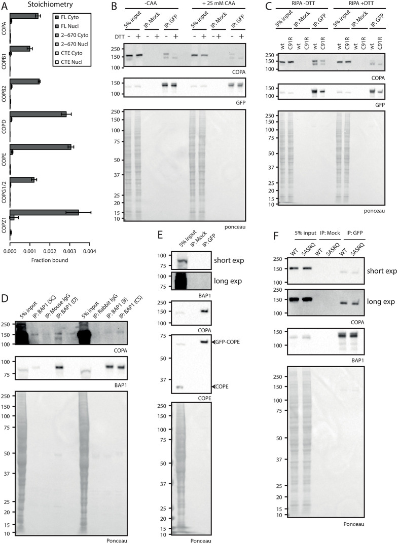
Fig 5. Characterization and validation of the BAP1 –COPI interaction.
(A). Relative binding of COPI complex subunits: stoichiometry based on label-free quantification of experiment in Fig 4. (B and C). Immunoblot analysis of GFP-BAP1 IP experiments. HeLa cells expressing GFP-BAP1 were grown and lysates were supplemented with CAA or DTT as indicated and used for mock or GFP immunoprecipitation. Blots were analyzed using listed antibodies. (D). BAP1 endogenous IPs using different BAP1 antibodies. HeLa FRT wt cell lysate was used for endogenous immunoprecipitation and blots were analyzed using listed antibodies. (E). Immunoblot analysis of GFP-COPE IP. Cell lysate of GFP-COPE expressing cells was used in GFP immunoprecipitation. Blots were analyzed using listed antibodies. (F). BAP1 C-terminal tail mutational analysis. Cell lysates of GFP-BAP1 wt or SASRQ c-terminally mutated BAP1 was used for mock or GFP immunoprecipitation. Blots were analyzed using listed antibodies.

