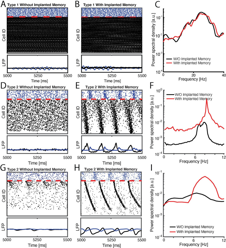
Fig 1. Type 2 model networks respond to sparse strengthening of excitatiory synapses or increasing excitatory input, through emergence of low-frequency rhythms and phase-locking.
SIMULATED DATA: Raster plots (top) and cumulative signal (bottom) generated by inibitory (blue), and excitatory (black) neurons in a wake-like (high-ACh) state before (A) and after (B) initial memory storage. C) Fourier transform of the wake-like excitatory network signal before (black) and after initial memory storage (red) reveal enhanced beta/gamma oscillations. (D, E) raster plots (top) and cumulative signal (bottom) of NREM-like (low-ACh) state before (D) and after (E) initial storage (i.e. 20 fold strengthening of recurrent synapses between pairs of 25% of excitatory engram neurons). F) Fourier transform of the NREM-like excitatory network signal before (black) and after initial memory storage (red) reveal enhanced slow oscillations in theta range. (G, H) raster plots (top) and cumulative signal (bottom) of NREM-like (low-ACh) state in a network with removed reciprocal excitatory synapses, before (G) and after (H) initial storage (i.e. 5% increase in the mean external drive, Iext, to excitatory engram neurons). I) Fourier transform of the NREM-like excitatory network signal before (black) and after initial memory storage (red) reveal enhanced slow oscillations in theta range. On all rasterplots neurons are order as a function of their frequency in wake-like state.

