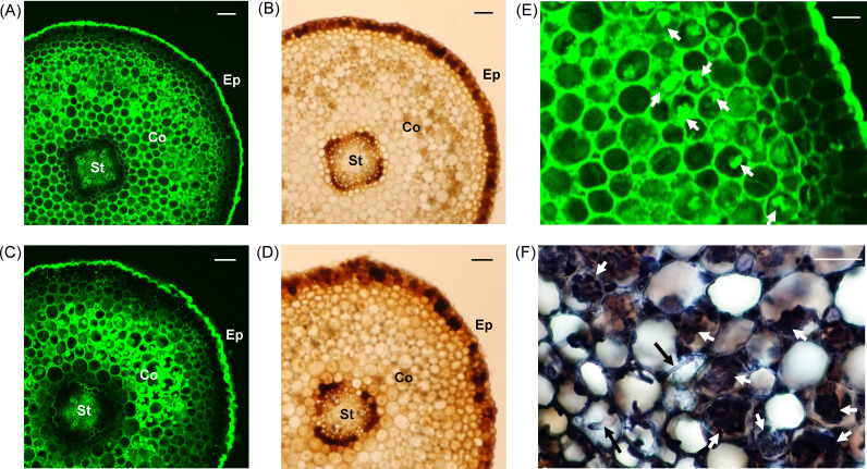
Fig 2. Zn localization in the roots of A. japonica.
(A) Zn localization in roots collected in July 2017 (summer). (B) Optical microscopically observed root sections collected in July 2017 (summer). (C) Zn localization in roots collected in January 2018 (winter). (D) Optical microscopically observed root sections collected in January 2018 (winter). (E) The extended images of the root cortex and epidermis of Fig 2A. (F) Trypan-blue stained fungal structures in the roots collected in July 2017. The green color indicates fluorescence derived from the Zinpyr-1 complex with Zn. St, stele; Co, cortex; Ep, epidermis. White arrows indicate the fungal structures, and black arrows indicate fungal hyphae. The Scale bars in Fig 2A–2D represent 100 μm, and Fig 2E and 2F represent 50 μm.

