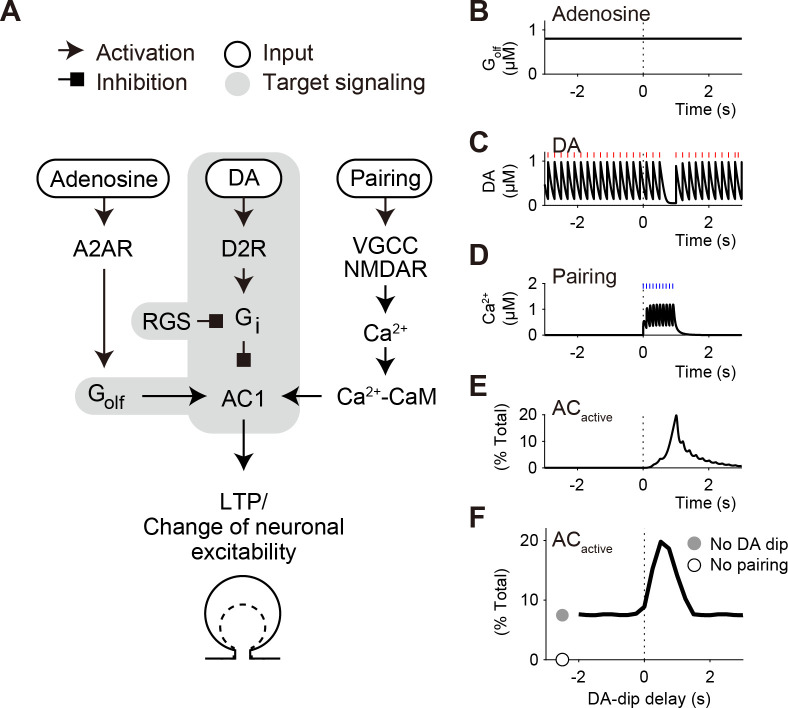
Fig 1. The D2 model performs the coincidence detection between DA dip and pre–post pairing.
(A) Three signaling cascades toward AC1 for LTP in D2 SPNs of the striatum [10]. First, adenosine stimulates A2AR, which produces Golf-GTP to activate AC1. Second, basal DA signal leads to the activity of D2R, and then produces a GTP from of Gi (Gi-GTP), which inhibits the AC1 activity. Third, pre–post pairing signal leads to the postsynaptic increase in Ca2+-CaM that stimulates AC1. Signaling in the gray shaded area is modeled in the present D2 model. (B-F) Coincidence detection between DA dip and pre–post pairing. (B) In Iino et al. (2020), A2AR were pharmacologically activated to give a continuous signal of Golf-GTP [1]. (C) DA fibers are optogenetically stimulated tonically at 5Hz (red lines). The tonic stimulation accompanies a 0.5-s pause (DA dip, tDA,delay = 0.5 s), as a representation of unexpected reward omission. (D) Sensory/action signals are represented by pre–post pairing at 10 times and 10 Hz (blue lines), which gives a transient Ca2+ signal. (E) The Golf, Gi, and Ca2+-CaM signals transiently activates AC1 (ACactive). (F) A timing window for DA-dip delay on the maximal amplitudes of ACactive. The D2 model is simulated under the non-competitive binding among Golf, Gi, and Ca2+-CaM (standard non-competitive model; see S1 Fig and section A in S1 Appendix).

