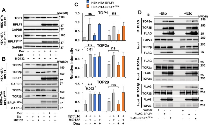
Fig 1. BPLF1 selectively binds to TOP2 and inhibits the degradation of TOP2 in cells treated with topoisomerase poisons.
HEK-293T cell expressing inducible FLAG-BPLF1 or FLAG-BPLF1C61A were seeded into 6 well plates and treated with 1.5 μg/ml Dox for 24 h. After treatment for 3 h with 5 μM of the TOP1 poison Camptothecin (Cpt) or 6 h with 40 μM of the TOP2 poison Etoposide (Eto) with or without the addition of 10 μM MG132, protein expression was analyzed in western blots probed with the indicated antibodies. GAPDH was used as the loading control. (A) Representative western blots illustrating the expression of TOP1 in control and Cpt treated cells. The proteasome-dependent degradation of TOP1 induced by the treatment was not affected by the expression of BPLF1 or BPLF1C61A in Dox treated cells. (B) Representative western blots illustrating the expression of TOP2α and TOP2β in Etoposide treated cells. Expression of BPLF1 protected TOP2α and TOP2β from Etoposide-induced proteasomal degradation while BPLF1C61A had no appreciable effect. (C) The intensity of the TOP1, TOP2α and TOP2β specific bands in 5 (TOP1) or 6 (TOP2α and TOP2β) independent experiments was quantified using the ImageJ software. The data are presented as intensity of the bands in Cpt/Eto treated samples relative to untreated control after normalization to the GAPDH loading control. Statistical analysis was performed using Student’s t-test. **P≤ 0.01; ns, not significant. (D) HEK293T cells transfected with FLAG-BPLF1, FLAG-BPLF1C61A, or FLAG-empty vector were treated with 40 μM Etoposide for 30 min and cell lysates were either immunoprecipitated with anti-FLAG conjugated agarose beads or incubated for 3 h with anti-TOP2α or TOP2β antibodies followed by the capture of immunocomplexes with protein-G coated beads. Catalytically active and inactive BPLF1 co-immunoprecipitate with both TOP2α and TOP2β in untreated and Etoposide treated cells (upper panels). Conversely, TOP2α (middle panels) and TOP2β (lower panels) interact with both catalytically active and inactive BPLF1. Representative western blots from one of two independent experiments where all conditions were tested in parallel are shown.

