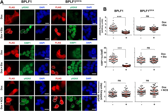
Fig 3. BPLF1 selectively inhibits the detection of TOP2-induced DNA damage.
HEK-rtTA-BPLF1/BPLF1C61A cells grown on cover-slides were treated with 1.5 μg/ml Dox for 24 h to induce the expression of BPLF1 followed by treatment for 1 h with 40 μM Etoposide or 0.5 μg/ml of the radiomimetic Neocarzinostatin (NCS) before staining with the indicated antibodies. (A) The cells were co-stained with antibodies against FLAG (red) and antibodies to γH2AX or 53BP1 (green) and the nuclei were stained with DAPI (blue). The expression of catalytically active BPLF1 was associated with a significant decrease of Etoposide induced nuclear γH2AX fluorescence and decreased formation of 53BP1 foci. BPLF1C61A had no effect. Neither the catalytically active nor the inactive BPLF1 affected the induction of γH2AX in cells treated with NCS. Representative micrographs from one of two experiments where all conditions were tested in parallel are shown. Scale bar = 10 μm. (B) Quantification of γH2AX fluorescence intensity and number of 53BP1 foci in BPLF1/BPLF1C61A positive and negative cells from the same images. The Mean ± SD of fluorescence intensity or number of dots in at least 50 BPLF1-positive and 50 BPLF1-negative cells recorded in each condition is shown. Statistical analysis was performed using Student’s t-test. ***P ≤0.001; ns, not significant.

