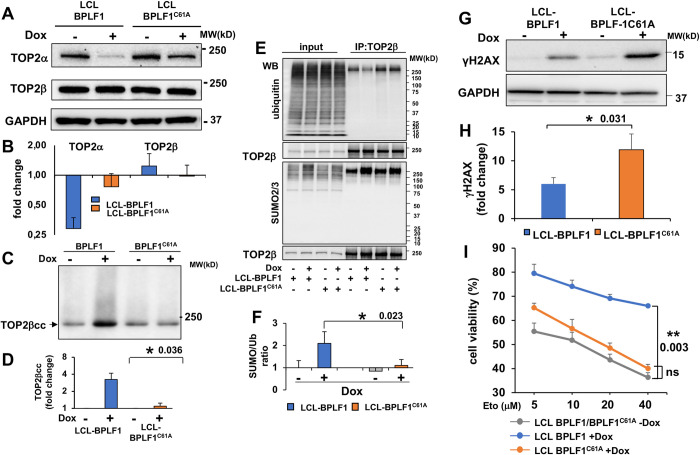
Fig 6. BPLF1 regulates the expression and activity of TOP2 in productively infected LCLs.
The productive virus cycle was induced by treatment with 1.5 μg/ml Dox in LCL cells carrying recombinant EBV encoding wild type or catalytic mutant BPLF1 and a tetracycline regulated BZLF1 transactivator. Induction of the productive cycle was associated with a highly reproducible downregulation of TOP2α while TOP2β was either unchanged or slightly increased. The effect was stronger in cells expressing wild type BPLF1. (A) Representative western blots illustrating the expression of TOP2α and TOP2β in control and induced cells. (B) The intensity of the specific bands was quantified using the ImageJ in three to five independent experiments and fold change in induced versus control cells was calculated after normalization to the GAPDH loading control. (C) The formation of TOP2βccs was investigated by RADAR assays in untreated and induced LCLs. Representative western blot illustrating the significant increase of TOP2ccs upon induction of the productive virus cycle in LCL cells expressing catalytically active BPLF1. BPLF1C61A had no appreciable effect. One representative western blot is shown. (D) Quantification of the intensity of the TOP2β smears in three independent experiments. Fold increase was calculated as the ratio between the smear intensity in control versus induced cells. *P≤0.05. (E) TOP2β was immunoprecipitated from total cell lysates of control and induced LCLs and western blots were probed with antibodies to TOP2β, ubiquitin and SUMO2/3. Western blots illustrating the increase of SUMOylated TOP2β in productively infected cells and selective decrease of ubiquitinated TOP2β in cells expressing catalytically active BPLF1. One representative experiment out of three is shown. (F) The intensity of the bands corresponding to immunoprecipitated TOP2β and ubiquitinated or SUMOylated species was quantified using the ImageJ software and the SUMO/Ub ratio was calculated after normalization to immunoprecipitated TOP2β. The mean ± SE of three independent experiments is shown. *P<0.05. (G) Representative western blot illustrating the expression of the DDR marker γH2AX in control and induced LCL-EBV-BPLF1/BPLF1C61A cells. (H) The intensity of γH2AX bands were quantified by densitometry in eight independent experiments. The fold increase in induced versus control cells was calculated after normalization to the GAPDH loading control and to the level of induction as assessed by the intensity of the BMRF1 specific band. Statistical analysis was performed using Student’s t-test. *P≤0.05. (I) The productive cycle was induced in LCL-EBV-BPLF1/BPLF1C61A by culture for 72 h in the presence 1.5 μg/ml Dox. After washing and counting, 5x10⁴ live cells were seeded in triplicate wells of 96 well plates and treated overnight with the indicated concentration of Etoposide before assessing cell viability by MTT assays. Catalytically active BPLF1 enhanced cell viability over a wide range of Etoposide concentration while BPLF1C61A had no appreciable effect. The mean ± SE of cell viability in three independent experiments is shown.**P≤0.01.

