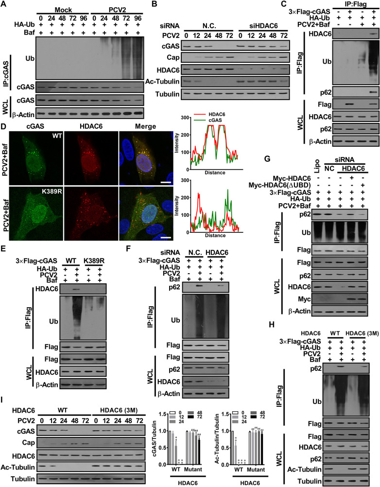
Fig 4. PCV2 infection activates HDAC6 to mediate the transport of poly-ubiquitin cGAS to the lysosome via HDAC6 mediation.
(A) PK-15 cells were infected with PCV2 (MOI = 1) for the indicated times and the poly-ubiquitination levels and protein levels of cGAS were analyzed. (B) PK-15 cells were transfected with HDAC6 specific siRNA (siHDAC6) or siRNA negative control (siN.C.) for 24 h before PCV2 infection (MOI = 5), and then the levels of porcine cGAS, PCV2 Cap, HDAC6, Ac-tubulin were determined by western blotting at the indicated time post-infection. (C) PK-15 cells were transfected plasmids indicated, then infected with PCV2 (MOI = 5) in the presence of Baf for 48 h. the interaction of ubiquitinated cGAS with HDAC6 and p62 was analyzed. (D) The cGAS-/- PK-15 cells transfected with Flag-cGAS, Flag-cGAS (K389R) expression constructs were infected with PCV2 in the presence of Baf. The colocalization of porcine cGAS and HDAC6 were observed under confocal microscopy. Scale bar, 10 μm. The co-localization signals of targeted proteins were analyzed as intensity profiles of indicated proteins along the plotted lines by Image J line scan analysis. (E) PK-15 cells were transfected expression vectors as indicated, then infected with PCV2 in the presence of Baf for 48 h. Subsequently, the ubiquitination levels of cGAS and cGAS (K389R), and their ability to bind HDAC6 were measured. (F) PK-15 cells were transfected with HDAC6 specific siRNA (siHDAC6) or siRNA negative control (siN.C.) and indicated plasmids for 24 h. Then these cells were infected with PCV2 (MOI = 5) or mock in the presence of Baf for 48 h; the interaction of ubiquitinated cGAS with P62 was analyzed. (G) PK-15 cells were transfected with HDAC6 specific siRNA (siHDAC6) or siRNA negative control (siN.C.) and indicated plasmids, meanwhile, Myc-HDAC6 (FL), Myc-HDAC6 (ΔUBD) were reconstituted in the cells transfected siHDAC6. Then, these cells were infected with PCV2 (MOI = 5) for 48 h, and the interaction of ubiquitinated cGAS with p62 was analyzed in these cells. (H) The HDAC6-/- PK-15 cells were transfected wild-type HDAC6 (WT) or mutated HDAC6-3M (D538A/D608A/S609A) for 24 h, then infected with PCV2 (MOI = 5) for another 48 h, and the interaction of ubiquitinated cGAS with p62 was analyzed. (I) The HDAC6-/- PK-15 cells transfected wild-type HDAC6 (WT) or mutated HDAC6-3M for 24 h, then infected with PCV2 (MOI = 5) for the indicated time, and then the levels of porcine cGAS, PCV2 Cap, and Ac-Tubulin were determined by western blotting. * P < 0.05, ** P < 0.01 (compared with infection at 0 h), # P < 0.05, ## P < 0.01 (compared with wild type HDAC6 group in indicated same time).

